 Movie
Movie Controller


+ Open data

- Basic information
| Entry | Database: PDB / ID: 6pib | |||||||||
|---|---|---|---|---|---|---|---|---|---|---|
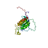
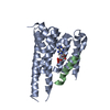
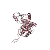
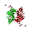
| Title | Structure of the Klebsiella pneumoniae LpxH-AZ1 complex | |||||||||
 Components Components | UDP-2,3-diacylglucosamine hydrolase | |||||||||
 Keywords Keywords | BIOSYNTHETIC PROTEIN / lipid A / LpxH / AZ1 / inhibitor | |||||||||
| Function / homology |  Function and homology information Function and homology informationUDP-2,3-diacylglucosamine diphosphatase / UDP-2,3-diacylglucosamine hydrolase activity / extrinsic component of plasma membrane / lipid A biosynthetic process / manganese ion binding / cytoplasm Similarity search - Function | |||||||||
| Biological species |  Klebsiella pneumoniae (bacteria) Klebsiella pneumoniae (bacteria) | |||||||||
| Method |  X-RAY DIFFRACTION / SYNCHROTRON / MOLECULAR REPLACEMENT / Resolution: 2.26 Å X-RAY DIFFRACTION / SYNCHROTRON / MOLECULAR REPLACEMENT / Resolution: 2.26 Å | |||||||||
 Authors Authors | Cho, J. / Zhou, P. | |||||||||
| Funding support |  United States, 2items United States, 2items
| |||||||||
 Citation Citation | Journal: Proc.Natl.Acad.Sci.USA / Year: 2020 Title: Structural basis of the UDP-diacylglucosamine pyrophosphohydrolase LpxH inhibition by sulfonyl piperazine antibiotics. Authors: Cho, J. / Lee, M. / Cochrane, C.S. / Webster, C.G. / Fenton, B.A. / Zhao, J. / Hong, J. / Zhou, P. | |||||||||
| History |
|
- Structure visualization
| Structure viewer | Molecule: MolmilJmol/JSmol |
|---|
- Downloads & links
-Download
| PDBx/mmCIF format | 6pib.cif.gz | 69.3 KB | Display | PDBx/mmCIF format |
|---|---|---|---|---|
| PDB format | pdb6pib.ent.gz | 47.8 KB | Display | PDB format |
| PDBx/mmJSON format | 6pib.json.gz | Tree view | PDBx/mmJSON format | |
| Others |  Other downloads Other downloads |
-Validation report
| Arichive directory | https://data.pdbj.org/pub/pdb/validation_reports/pi/6pibftp://data.pdbj.org/pub/pdb/validation_reports/pi/6pib | HTTPS FTP |
|---|
-Related structure data
| Related structure data |  6ph9C  6pj3C  5k8kS S: Starting model for refinement C: citing same article ( |
|---|---|
| Similar structure data |


-Links
 PDBj
PDBj
- Assembly
| Deposited unit | 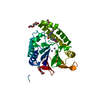
| ||||||||
|---|---|---|---|---|---|---|---|---|---|
| 1 |
| ||||||||
| Unit cell |
|
-Components
| #1: Protein | Mass: 29726.809 Da / Num. of mol.: 1 Source method: isolated from a genetically manipulated source Source: (gene. exp.) Klebsiella pneumoniae (bacteria)Gene: lpxH_1, lpxH, C3483_19950, NCTC9128_00880, SAMEA104305404_03891 Production host: References: UniProt: A0A1S0WIC1, UniProt: A6T5R0*PLUS, UDP-2,3-diacylglucosamine diphosphatase | ||||||||
|---|---|---|---|---|---|---|---|---|---|
| #2: Chemical |   Mass: 54.938 Da / Num. of mol.: 2 / Source method: obtained synthetically / Formula: Mn Mass: 54.938 Da / Num. of mol.: 2 / Source method: obtained synthetically / Formula: Mn#3: Chemical | ChemComp-OKV / |   Mass: 453.478 Da / Num. of mol.: 1 / Source method: obtained synthetically / Formula: C21H22F3N3O3S / Feature type: SUBJECT OF INVESTIGATION Mass: 453.478 Da / Num. of mol.: 1 / Source method: obtained synthetically / Formula: C21H22F3N3O3S / Feature type: SUBJECT OF INVESTIGATION#4: Chemical |   Mass: 194.226 Da / Num. of mol.: 3 / Source method: obtained synthetically / Formula: C8H18O5 / Comment: precipitant*YM Mass: 194.226 Da / Num. of mol.: 3 / Source method: obtained synthetically / Formula: C8H18O5 / Comment: precipitant*YM#5: Water | ChemComp-HOH / |  Mass: 18.015 Da / Num. of mol.: 46 / Source method: isolated from a natural source / Formula: H2O Mass: 18.015 Da / Num. of mol.: 46 / Source method: isolated from a natural source / Formula: H2OHas ligand of interest | Y | |
-Experimental details
-Experiment
| Experiment | Method: X-RAY DIFFRACTION / Number of used crystals: 1 |
|---|
- Sample preparation
| Crystal | Density Matthews: 2.93 Å3/Da / Density % sol: 58.03 % |
|---|---|
| Crystal grow | Temperature: 293 K / Method: vapor diffusion, sitting drop Details: 10 mM MES, pH 6.0, 100 mM NaCl, 0.5 mM DTT, 2.5% glycerol, 0.75% DMSO 25 mM sodium chloride, 10 mM magnesium chloride hexahydrate, 50 mM sodium citrate, pH 6.0, and 11% PEG 400. |
-Data collection
| Diffraction | Mean temperature: 100 K / Serial crystal experiment: N |
|---|---|
| Diffraction source | Source: SYNCHROTRON / Site: APS / Beamline: 24-ID-C / Wavelength: 0.9791 Å |
| Detector | Type: DECTRIS PILATUS 6M-F / Detector: PIXEL / Date: Jun 24, 2018 |
| Radiation | Protocol: SINGLE WAVELENGTH / Monochromatic (M) / Laue (L): M / Scattering type: x-ray |
| Radiation wavelength | Wavelength: 0.9791 Å / Relative weight: 1 |
| Reflection | Resolution: 2.26→45.96 Å / Num. obs: 16565 / % possible obs: 99.9 % / Redundancy: 9.9 % / Net I/σ(I): 20.32 |
| Reflection shell | Resolution: 2.26→2.34 Å / Num. unique obs: 1646 |
- Processing
| Software |
| |||||||||||||||||||||||||||||||||||||||||||||||||
|---|---|---|---|---|---|---|---|---|---|---|---|---|---|---|---|---|---|---|---|---|---|---|---|---|---|---|---|---|---|---|---|---|---|---|---|---|---|---|---|---|---|---|---|---|---|---|---|---|---|---|
| Refinement | Method to determine structure: MOLECULAR REPLACEMENT Starting model: 5k8k Resolution: 2.26→45.96 Å / SU ML: 0.23 / Cross valid method: FREE R-VALUE / σ(F): 1.37 / Phase error: 29.25
| |||||||||||||||||||||||||||||||||||||||||||||||||
| Solvent computation | Shrinkage radii: 0.9 Å / VDW probe radii: 1.11 Å | |||||||||||||||||||||||||||||||||||||||||||||||||
| Refinement step | Cycle: LAST / Resolution: 2.26→45.96 Å
| |||||||||||||||||||||||||||||||||||||||||||||||||
| Refine LS restraints |
| |||||||||||||||||||||||||||||||||||||||||||||||||
| LS refinement shell |
|
