 Movie
Movie Controller

[English] 日本語
 Yorodumi
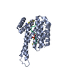
- PDB-6a5s: Structure of 14-3-3 gamma in complex with TFEB 14-3-3 binding motif -
+ Open data

- Basic information
| Entry | Database: PDB / ID: 6a5s | |||||||||
|---|---|---|---|---|---|---|---|---|---|---|
| Title | Structure of 14-3-3 gamma in complex with TFEB 14-3-3 binding motif | |||||||||
 Components Components |
| |||||||||
 Keywords Keywords | TRANSCRIPTION / PHOSPHOSERIN / REGULATION / TRANSCRIPTION FACTOR | |||||||||
| Function / homology |  Function and homology information Function and homology informationpositive regulation of cell-cell adhesion / phosphorylation-dependent protein binding / positive regulation of T cell mediated immune response to tumor cell / regulation of neuron differentiation / protein kinase C inhibitor activity / Regulation of localization of FOXO transcription factors / SPOP-mediated proteasomal degradation of PD-L1(CD274) / Activation of BAD and translocation to mitochondria / regulation of signal transduction / SARS-CoV-2 targets host intracellular signalling and regulatory pathways ...positive regulation of cell-cell adhesion / phosphorylation-dependent protein binding / positive regulation of T cell mediated immune response to tumor cell / regulation of neuron differentiation / protein kinase C inhibitor activity / Regulation of localization of FOXO transcription factors / SPOP-mediated proteasomal degradation of PD-L1(CD274) / Activation of BAD and translocation to mitochondria / regulation of signal transduction / SARS-CoV-2 targets host intracellular signalling and regulatory pathways / negative regulation of protein kinase activity / cellular response to glucose starvation / Chk1/Chk2(Cds1) mediated inactivation of Cyclin B:Cdk1 complex / SARS-CoV-1 targets host intracellular signalling and regulatory pathways / RHO GTPases activate PKNs / insulin-like growth factor receptor binding / negative regulation of TORC1 signaling / Loss of Nlp from mitotic centrosomes / Loss of proteins required for interphase microtubule organization from the centrosome / Transcriptional and post-translational regulation of MITF-M expression and activity / Recruitment of mitotic centrosome proteins and complexes / Recruitment of NuMA to mitotic centrosomes / Anchoring of the basal body to the plasma membrane / AURKA Activation by TPX2 / protein kinase C binding / TP53 Regulates Metabolic Genes / Translocation of SLC2A4 (GLUT4) to the plasma membrane / protein sequestering activity / receptor tyrosine kinase binding / regulation of synaptic plasticity / positive regulation of T cell activation / cellular response to insulin stimulus / intracellular protein localization / Regulation of PLK1 Activity at G2/M Transition / regulation of protein localization / presynapse / mitochondrial matrix / protein domain specific binding / focal adhesion / signal transduction / RNA binding / extracellular exosome / membrane / identical protein binding / nucleus / cytosol / cytoplasm Similarity search - Function | |||||||||
| Biological species |  Homo sapiens (human) Homo sapiens (human) | |||||||||
| Method |  X-RAY DIFFRACTION / SYNCHROTRON / MOLECULAR REPLACEMENT / Resolution: 2.099 Å X-RAY DIFFRACTION / SYNCHROTRON / MOLECULAR REPLACEMENT / Resolution: 2.099 Å | |||||||||
 Authors Authors | Xu, Y. / Ren, J.Q. / Feng, W. | |||||||||
 Citation Citation | Journal: Autophagy / Year: 2019 Title: YWHA/14-3-3 proteins recognize phosphorylated TFEB by a noncanonical mode for controlling TFEB cytoplasmic localization. Authors: Xu, Y. / Ren, J. / He, X. / Chen, H. / Wei, T. / Feng, W. | |||||||||
| History |
|
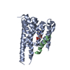
- Structure visualization
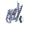
| Structure viewer | Molecule: MolmilJmol/JSmol |
|---|
- Downloads & links
-Download
| PDBx/mmCIF format | 6a5s.cif.gz | 220.5 KB | Display | PDBx/mmCIF format |
|---|---|---|---|---|
| PDB format | pdb6a5s.ent.gz | 175 KB | Display | PDB format |
| PDBx/mmJSON format | 6a5s.json.gz | Tree view | PDBx/mmJSON format | |
| Others |  Other downloads Other downloads |
-Validation report
| Arichive directory | https://data.pdbj.org/pub/pdb/validation_reports/a5/6a5sftp://data.pdbj.org/pub/pdb/validation_reports/a5/6a5s | HTTPS FTP |
|---|
-Related structure data
| Related structure data | 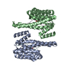 6a5qC  3uzdS S: Starting model for refinement C: citing same article ( |
|---|---|
| Similar structure data |


-Links
 PDBj
PDBj


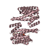
- Assembly
| Deposited unit | 
| ||||||||
|---|---|---|---|---|---|---|---|---|---|
| 1 | 
| ||||||||
| 2 | 
| ||||||||
| Unit cell |
|
-Components
| #1: Protein | Mass: 28423.668 Da / Num. of mol.: 4 Source method: isolated from a genetically manipulated source Source: (gene. exp.) Homo sapiens (human) / Gene: YWHAG / Production host:  #2: Protein/peptide | Mass: 1557.616 Da / Num. of mol.: 4 / Source method: obtained synthetically / Source: (synth.) Homo sapiens (human)#3: Chemical | ChemComp-NA / |   Mass: 22.990 Da / Num. of mol.: 1 / Source method: obtained synthetically / Formula: Na Mass: 22.990 Da / Num. of mol.: 1 / Source method: obtained synthetically / Formula: Na#4: Chemical | ChemComp-MG / |   Mass: 24.305 Da / Num. of mol.: 1 / Source method: obtained synthetically / Formula: Mg Mass: 24.305 Da / Num. of mol.: 1 / Source method: obtained synthetically / Formula: Mg#5: Water | ChemComp-HOH / |  Mass: 18.015 Da / Num. of mol.: 803 / Source method: isolated from a natural source / Formula: H2O Mass: 18.015 Da / Num. of mol.: 803 / Source method: isolated from a natural source / Formula: H2OHas protein modification | Y | |
|---|
-Experimental details
-Experiment
| Experiment | Method: X-RAY DIFFRACTION / Number of used crystals: 1 |
|---|
- Sample preparation
| Crystal | Density Matthews: 2.66 Å3/Da / Density % sol: 53.77 % |
|---|---|
| Crystal grow | Temperature: 289 K / Method: evaporation / pH: 7.3 Details: 0.1 M Tris-HCl, 0.2 M calcium acetate, and 20% (w/v) PEG 3350 |
-Data collection
| Diffraction | Mean temperature: 100 K |
|---|---|
| Diffraction source | Source: SYNCHROTRON / Site: SSRF  / Beamline: BL17U / Wavelength: 0.9789 Å / Beamline: BL17U / Wavelength: 0.9789 Å |
| Detector | Type: ADSC QUANTUM 315 / Detector: CCD / Date: Dec 12, 2017 |
| Radiation | Protocol: SINGLE WAVELENGTH / Monochromatic (M) / Laue (L): M / Scattering type: x-ray |
| Radiation wavelength | Wavelength: 0.9789 Å / Relative weight: 1 |
| Reflection | Resolution: 2.099→50 Å / Num. obs: 73457 / % possible obs: 99 % / Redundancy: 5.7 % / Rmerge(I) obs: 0.142 / Net I/σ(I): 13.1 |
| Reflection shell | Resolution: 2.099→2.14 Å / Rmerge(I) obs: 0.664 / Mean I/σ(I) obs: 2.9 / Num. unique obs: 3618 / % possible all: 100 |
- Processing
| Software |
| |||||||||||||||||||||||||||||||||||||||||||||||||||||||||||||||||||||||||||||||||||||||||||||||||||||||||
|---|---|---|---|---|---|---|---|---|---|---|---|---|---|---|---|---|---|---|---|---|---|---|---|---|---|---|---|---|---|---|---|---|---|---|---|---|---|---|---|---|---|---|---|---|---|---|---|---|---|---|---|---|---|---|---|---|---|---|---|---|---|---|---|---|---|---|---|---|---|---|---|---|---|---|---|---|---|---|---|---|---|---|---|---|---|---|---|---|---|---|---|---|---|---|---|---|---|---|---|---|---|---|---|---|---|---|
| Refinement | Method to determine structure: MOLECULAR REPLACEMENT Starting model: 3UZD Resolution: 2.099→44.044 Å / SU ML: 0.21 / Cross valid method: THROUGHOUT / σ(F): 1.35 / Phase error: 25.67
| |||||||||||||||||||||||||||||||||||||||||||||||||||||||||||||||||||||||||||||||||||||||||||||||||||||||||
| Solvent computation | Shrinkage radii: 0.9 Å / VDW probe radii: 1.11 Å | |||||||||||||||||||||||||||||||||||||||||||||||||||||||||||||||||||||||||||||||||||||||||||||||||||||||||
| Displacement parameters | Biso max: 87.08 Å2 / Biso mean: 25.3333 Å2 / Biso min: 8.95 Å2 | |||||||||||||||||||||||||||||||||||||||||||||||||||||||||||||||||||||||||||||||||||||||||||||||||||||||||
| Refinement step | Cycle: final / Resolution: 2.099→44.044 Å
| |||||||||||||||||||||||||||||||||||||||||||||||||||||||||||||||||||||||||||||||||||||||||||||||||||||||||
| Refine LS restraints |
| |||||||||||||||||||||||||||||||||||||||||||||||||||||||||||||||||||||||||||||||||||||||||||||||||||||||||
| LS refinement shell | Refine-ID: X-RAY DIFFRACTION / Rfactor Rfree error: 0 / Total num. of bins used: 14
|
