 Movie
Movie Controller

[English] 日本語
 Yorodumi
- PDB-6ph9: Crystal Structure of the Klebsiella pneumoniae LpxH-lipid X complex -
+ Open data

- Basic information
| Entry | Database: PDB / ID: 6ph9 | |||||||||
|---|---|---|---|---|---|---|---|---|---|---|
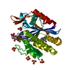
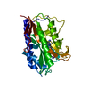
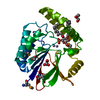
| Title | Crystal Structure of the Klebsiella pneumoniae LpxH-lipid X complex | |||||||||
 Components Components | UDP-2,3-diacylglucosamine hydrolase | |||||||||
 Keywords Keywords | BIOSYNTHETIC PROTEIN / lipid A / LpxH / lipid X | |||||||||
| Function / homology |  Function and homology information Function and homology informationUDP-2,3-diacylglucosamine diphosphatase / UDP-2,3-diacylglucosamine hydrolase activity / extrinsic component of plasma membrane / lipid A biosynthetic process / manganese ion binding / cytoplasm Similarity search - Function | |||||||||
| Biological species |  Klebsiella pneumoniae (bacteria) Klebsiella pneumoniae (bacteria) | |||||||||
| Method |  X-RAY DIFFRACTION / SYNCHROTRON / MOLECULAR REPLACEMENT / Resolution: 1.92 Å X-RAY DIFFRACTION / SYNCHROTRON / MOLECULAR REPLACEMENT / Resolution: 1.92 Å | |||||||||
 Authors Authors | Cho, J. / Zhou, P. | |||||||||
| Funding support |  United States, 2items United States, 2items
| |||||||||
 Citation Citation | Journal: Proc.Natl.Acad.Sci.USA / Year: 2020 Title: Structural basis of the UDP-diacylglucosamine pyrophosphohydrolase LpxH inhibition by sulfonyl piperazine antibiotics. Authors: Cho, J. / Lee, M. / Cochrane, C.S. / Webster, C.G. / Fenton, B.A. / Zhao, J. / Hong, J. / Zhou, P. | |||||||||
| History |
|
- Structure visualization
| Structure viewer | Molecule: MolmilJmol/JSmol |
|---|
- Downloads & links
-Download
| PDBx/mmCIF format | 6ph9.cif.gz | 76.6 KB | Display | PDBx/mmCIF format |
|---|---|---|---|---|
| PDB format | pdb6ph9.ent.gz | 52.6 KB | Display | PDB format |
| PDBx/mmJSON format | 6ph9.json.gz | Tree view | PDBx/mmJSON format | |
| Others |  Other downloads Other downloads |
-Validation report
| Arichive directory | https://data.pdbj.org/pub/pdb/validation_reports/ph/6ph9ftp://data.pdbj.org/pub/pdb/validation_reports/ph/6ph9 | HTTPS FTP |
|---|
-Related structure data
| Related structure data |  6pibC  6pj3C  5k8kS S: Starting model for refinement C: citing same article ( |
|---|---|
| Similar structure data |


-Links
 PDBj
PDBj
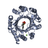
- Assembly
| Deposited unit | 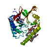
| ||||||||
|---|---|---|---|---|---|---|---|---|---|
| 1 |
| ||||||||
| Unit cell |
| ||||||||
| Components on special symmetry positions |
|
-Components
-Protein , 1 types, 1 molecules A
| #1: Protein | Mass: 29726.809 Da / Num. of mol.: 1 Source method: isolated from a genetically manipulated source Source: (gene. exp.) Klebsiella pneumoniae (bacteria)Gene: lpxH_1, lpxH, C3483_19950, NCTC9128_00880, SAMEA104305404_03891 Production host: References: UniProt: A0A1S0WIC1, UniProt: A6T5R0*PLUS, UDP-2,3-diacylglucosamine diphosphatase |
|---|
-Non-polymers , 6 types, 214 molecules 


| #2: Chemical | ChemComp-MN /  Mass: 54.938 Da / Num. of mol.: 4 / Source method: obtained synthetically / Formula: Mn Mass: 54.938 Da / Num. of mol.: 4 / Source method: obtained synthetically / Formula: Mn#3: Chemical | ChemComp-LP5 / ( |  Mass: 711.861 Da / Num. of mol.: 1 / Source method: obtained synthetically / Formula: C34H66NO12P / Feature type: SUBJECT OF INVESTIGATION Mass: 711.861 Da / Num. of mol.: 1 / Source method: obtained synthetically / Formula: C34H66NO12P / Feature type: SUBJECT OF INVESTIGATION#4: Chemical |  Mass: 194.226 Da / Num. of mol.: 2 / Source method: obtained synthetically / Formula: C8H18O5 / Comment: precipitant*YM Mass: 194.226 Da / Num. of mol.: 2 / Source method: obtained synthetically / Formula: C8H18O5 / Comment: precipitant*YM#5: Chemical | ChemComp-CA / |  Mass: 40.078 Da / Num. of mol.: 1 / Source method: obtained synthetically / Formula: Ca Mass: 40.078 Da / Num. of mol.: 1 / Source method: obtained synthetically / Formula: Ca#6: Chemical | ChemComp-EDO / |  Mass: 62.068 Da / Num. of mol.: 1 / Source method: isolated from a natural source / Formula: C2H6O2 Mass: 62.068 Da / Num. of mol.: 1 / Source method: isolated from a natural source / Formula: C2H6O2#7: Water | ChemComp-HOH / | Mass: 18.015 Da / Num. of mol.: 205 / Source method: isolated from a natural source / Formula: H2O |
|---|
-Details
| Has ligand of interest | Y |
|---|
-Experimental details
-Experiment
| Experiment | Method: X-RAY DIFFRACTION / Number of used crystals: 1 |
|---|
- Sample preparation
| Crystal | Density Matthews: 2.87 Å3/Da / Density % sol: 57.11 % |
|---|---|
| Crystal grow | Temperature: 293 K / Method: vapor diffusion, sitting drop Details: 4 mg/mL protein, 10 mM MES, pH 6.0, 100 mM NaCl, 0.5 mM DTT, 2.5% glycerol, 100mM calcium chloride dihydrate, 50mM HEPES, pH 7.0, 16.5% PEG 400 |
-Data collection
| Diffraction | Mean temperature: 100 K / Serial crystal experiment: N |
|---|---|
| Diffraction source | Source: SYNCHROTRON / Site: APS / Beamline: 24-ID-E / Wavelength: 0.97918 Å |
| Detector | Type: DECTRIS EIGER X 16M / Detector: PIXEL / Date: Aug 14, 2018 |
| Radiation | Protocol: SINGLE WAVELENGTH / Monochromatic (M) / Laue (L): M / Scattering type: x-ray |
| Radiation wavelength | Wavelength: 0.97918 Å / Relative weight: 1 |
| Reflection | Resolution: 1.92→45.895 Å / Num. obs: 26262 / % possible obs: 99.94 % / Redundancy: 19.7 % / Net I/σ(I): 33.03 |
| Reflection shell | Resolution: 1.92→1.989 Å / Num. unique obs: 2588 |
- Processing
| Software |
| ||||||||||||||||||||||||||||||||||||||||||||||||||||||||||||||||||||||
|---|---|---|---|---|---|---|---|---|---|---|---|---|---|---|---|---|---|---|---|---|---|---|---|---|---|---|---|---|---|---|---|---|---|---|---|---|---|---|---|---|---|---|---|---|---|---|---|---|---|---|---|---|---|---|---|---|---|---|---|---|---|---|---|---|---|---|---|---|---|---|---|
| Refinement | Method to determine structure: MOLECULAR REPLACEMENT Starting model: 5K8K Resolution: 1.92→45.895 Å / SU ML: 0.25 / Cross valid method: FREE R-VALUE / σ(F): 1.38 / Phase error: 21.91
| ||||||||||||||||||||||||||||||||||||||||||||||||||||||||||||||||||||||
| Solvent computation | Shrinkage radii: 0.9 Å / VDW probe radii: 1.11 Å | ||||||||||||||||||||||||||||||||||||||||||||||||||||||||||||||||||||||
| Refinement step | Cycle: LAST / Resolution: 1.92→45.895 Å
| ||||||||||||||||||||||||||||||||||||||||||||||||||||||||||||||||||||||
| Refine LS restraints |
| ||||||||||||||||||||||||||||||||||||||||||||||||||||||||||||||||||||||
| LS refinement shell |
|
