 Movie
Movie Controller

[English] 日本語
 Yorodumi
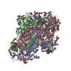
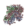
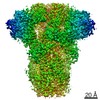
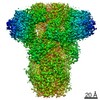
- PDB-6nzk: Structural basis for human coronavirus attachment to sialic acid ... -
+ Open data

- Basic information
| Entry | Database: PDB / ID: 6nzk | |||||||||
|---|---|---|---|---|---|---|---|---|---|---|
| Title | Structural basis for human coronavirus attachment to sialic acid receptors | |||||||||
 Components Components | Spike surface glycoprotein | |||||||||
 Keywords Keywords | VIRAL PROTEIN / Coronavirus spike glycoprotein / sialic acid / HCoV-OC43 / fusion protein | |||||||||
| Function / homology |  Function and homology information Function and homology informationhost cell endoplasmic reticulum-Golgi intermediate compartment membrane / receptor-mediated virion attachment to host cell / endocytosis involved in viral entry into host cell / fusion of virus membrane with host plasma membrane / fusion of virus membrane with host endosome membrane / viral envelope / host cell plasma membrane / virion membrane / membrane Similarity search - Function | |||||||||
| Biological species |  Human coronavirus OC43 Human coronavirus OC43 | |||||||||
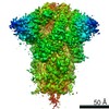
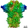
| Method | ELECTRON MICROSCOPY / single particle reconstruction / cryo EM / Resolution: 2.8 Å | |||||||||
 Authors Authors | Tortorici, M.A. / Walls, A.C. / Lang, Y. / Wang, C. / Li, Z. / Koerhuis, D. / Boons, G.J. / Bosch, B.J. / Rey, F.A. / de Groot, R. / Veesler, D. | |||||||||
| Funding support |  United States, 1items United States, 1items
| |||||||||
 Citation Citation | Journal: Nat Struct Mol Biol / Year: 2019 Title: Structural basis for human coronavirus attachment to sialic acid receptors. Authors: M Alejandra Tortorici / Alexandra C Walls / Yifei Lang / Chunyan Wang / Zeshi Li / Danielle Koerhuis / Geert-Jan Boons / Berend-Jan Bosch / Félix A Rey / Raoul J de Groot / David Veesler /   Abstract: Coronaviruses cause respiratory tract infections in humans and outbreaks of deadly pneumonia worldwide. Infections are initiated by the transmembrane spike (S) glycoprotein, which binds to host ...Coronaviruses cause respiratory tract infections in humans and outbreaks of deadly pneumonia worldwide. Infections are initiated by the transmembrane spike (S) glycoprotein, which binds to host receptors and fuses the viral and cellular membranes. To understand the molecular basis of coronavirus attachment to oligosaccharide receptors, we determined cryo-EM structures of coronavirus OC43 S glycoprotein trimer in isolation and in complex with a 9-O-acetylated sialic acid. We show that the ligand binds with fast kinetics to a surface-exposed groove and that interactions at the identified site are essential for S-mediated viral entry into host cells, but free monosaccharide does not trigger fusogenic conformational changes. The receptor-interacting site is conserved in all coronavirus S glycoproteins that engage 9-O-acetyl-sialogycans, with an architecture similar to those of the ligand-binding pockets of coronavirus hemagglutinin esterases and influenza virus C/D hemagglutinin-esterase fusion glycoproteins. Our results demonstrate these viruses evolved similar strategies to engage sialoglycans at the surface of target cells. | |||||||||
| History |
|
- Structure visualization
| Movie |
Movie viewer |
|---|---|
| Structure viewer | Molecule: MolmilJmol/JSmol |
- Downloads & links
-Download
| PDBx/mmCIF format | 6nzk.cif.gz | 723.1 KB | Display | PDBx/mmCIF format |
|---|---|---|---|---|
| PDB format | pdb6nzk.ent.gz | 588.3 KB | Display | PDB format |
| PDBx/mmJSON format | 6nzk.json.gz | Tree view | PDBx/mmJSON format | |
| Others |  Other downloads Other downloads |
-Validation report
| Arichive directory | https://data.pdbj.org/pub/pdb/validation_reports/nz/6nzkftp://data.pdbj.org/pub/pdb/validation_reports/nz/6nzk | HTTPS FTP |
|---|
-Related structure data
| Related structure data |  0557MC  6ohwC M: map data used to model this data C: citing same article ( |
|---|---|
| Similar structure data |


-Links
 PDBj
PDBj- Assembly
| Deposited unit | 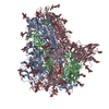
|
|---|---|
| 1 |
|
-Components
-Protein / Non-polymers , 2 types, 399 molecules ABC
| #1: Protein | Mass: 146438.312 Da / Num. of mol.: 3 / Mutation: R754G,R755G,R757G Source method: isolated from a genetically manipulated source Source: (gene. exp.) Human coronavirus OC43 / Production host:  Homo sapiens (human) / References: UniProt: Q696P8 Homo sapiens (human) / References: UniProt: Q696P8#7: Water | ChemComp-HOH / | Mass: 18.015 Da / Num. of mol.: 396 / Source method: isolated from a natural source / Formula: H2O |
|---|
-Sugars , 5 types, 48 molecules 
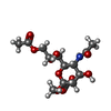
| #2: Polysaccharide | beta-D-mannopyranose-(1-4)-2-acetamido-2-deoxy-beta-D-glucopyranose-(1-4)-2-acetamido-2-deoxy-beta- ...beta-D-mannopyranose-(1-4)-2-acetamido-2-deoxy-beta-D-glucopyranose-(1-4)-2-acetamido-2-deoxy-beta-D-glucopyranose #3: Polysaccharide | alpha-D-mannopyranose-(1-3)-beta-D-mannopyranose-(1-4)-2-acetamido-2-deoxy-beta-D-glucopyranose-(1- ...alpha-D-mannopyranose-(1-3)-beta-D-mannopyranose-(1-4)-2-acetamido-2-deoxy-beta-D-glucopyranose-(1-4)-2-acetamido-2-deoxy-beta-D-glucopyranose #4: Polysaccharide | 2-acetamido-2-deoxy-beta-D-glucopyranose-(1-4)-2-acetamido-2-deoxy-beta-D-glucopyranose #5: Sugar | ChemComp-NAG /  Type: D-saccharide, beta linking / Mass: 221.208 Da / Num. of mol.: 15 / Source method: isolated from a natural source / Formula: C8H15NO6 Type: D-saccharide, beta linking / Mass: 221.208 Da / Num. of mol.: 15 / Source method: isolated from a natural source / Formula: C8H15NO6#6: Sugar |  Type: D-saccharide / Mass: 365.333 Da / Num. of mol.: 3 / Source method: isolated from a natural source / Formula: C14H23NO10 Type: D-saccharide / Mass: 365.333 Da / Num. of mol.: 3 / Source method: isolated from a natural source / Formula: C14H23NO10 |
|---|
-Details
| Has protein modification | Y |
|---|
-Experimental details
-Experiment
| Experiment | Method: ELECTRON MICROSCOPY |
|---|---|
| EM experiment | Aggregation state: PARTICLE / 3D reconstruction method: single particle reconstruction |
- Sample preparation
| Component | Name: HCoV-OC43 spike glycoprotein ectodomain in complex with 9-O-acetyl sialic acid Type: COMPLEX / Entity ID: #1 / Source: RECOMBINANT |
|---|---|
| Molecular weight | Experimental value: NO |
| Source (natural) | Organism: Betacoronavirus |
| Source (recombinant) | Organism: Homo sapiens (human) |
| Buffer solution | pH: 8 |
| Specimen | Embedding applied: NO / Shadowing applied: NO / Staining applied: NO / Vitrification applied: YES |
| Specimen support | Grid type: C-flat-1.2/1.3 4C |
| Vitrification | Cryogen name: ETHANE |
- Electron microscopy imaging
| Experimental equipment |  Model: Titan Krios / Image courtesy: FEI Company |
|---|---|
| Microscopy | Model: FEI TITAN KRIOS |
| Electron gun | Electron source:  FIELD EMISSION GUN / Accelerating voltage: 300 kV / Illumination mode: FLOOD BEAM FIELD EMISSION GUN / Accelerating voltage: 300 kV / Illumination mode: FLOOD BEAM |
| Electron lens | Mode: BRIGHT FIELD |
| Image recording | Electron dose: 70 e/Å2 / Detector mode: COUNTING / Film or detector model: GATAN K2 SUMMIT (4k x 4k) |
- Processing
| EM software |
| ||||||||||||||||||||
|---|---|---|---|---|---|---|---|---|---|---|---|---|---|---|---|---|---|---|---|---|---|
| CTF correction | Type: PHASE FLIPPING AND AMPLITUDE CORRECTION | ||||||||||||||||||||
| Symmetry | Point symmetry: C3 (3 fold cyclic) | ||||||||||||||||||||
| 3D reconstruction | Resolution: 2.8 Å / Resolution method: FSC 0.143 CUT-OFF / Num. of particles: 178356 / Symmetry type: POINT |
