 Movie
Movie Controller

[English] 日本語
 Yorodumi
- EMDB-6369: Structure of full-length IP3R1 channel in the apo-state determine... -
+ Open data

- Basic information
| Entry | Database: EMDB / ID: EMD-6369 | |||||||||
|---|---|---|---|---|---|---|---|---|---|---|
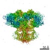
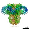
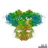
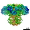
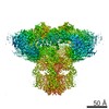
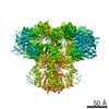
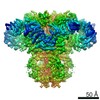
| Title | Structure of full-length IP3R1 channel in the apo-state determined by single particle cryo-EM | |||||||||
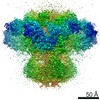
 Map data Map data | Reconstruction of IP3R1/Calcium release channel from the rat cerebellum | |||||||||
 Sample Sample |
| |||||||||
 Keywords Keywords | inositol 1 / 4 / 5-trisphosphate receptor / calcium release channel / single-particle cryo-EM / calcium signaling / 3D reconstruction | |||||||||
| Function / homology |  Function and homology information Function and homology informationEffects of PIP2 hydrolysis / Antigen activates B Cell Receptor (BCR) leading to generation of second messengers / inositol 1,4,5-trisphosphate receptor activity involved in regulation of postsynaptic cytosolic calcium levels / release of sequestered calcium ion into cytosol by endoplasmic reticulum / cGMP effects / smooth endoplasmic reticulum membrane / platelet dense granule membrane / Elevation of cytosolic Ca2+ levels / platelet dense tubular network / calcineurin complex ...Effects of PIP2 hydrolysis / Antigen activates B Cell Receptor (BCR) leading to generation of second messengers / inositol 1,4,5-trisphosphate receptor activity involved in regulation of postsynaptic cytosolic calcium levels / release of sequestered calcium ion into cytosol by endoplasmic reticulum / cGMP effects / smooth endoplasmic reticulum membrane / platelet dense granule membrane / Elevation of cytosolic Ca2+ levels / platelet dense tubular network / calcineurin complex / epithelial fluid transport / ion channel modulating, G protein-coupled receptor signaling pathway / phospholipase C-activating G protein-coupled acetylcholine receptor signaling pathway / inositol 1,4,5-trisphosphate-gated calcium channel activity / calcium import into the mitochondrion / voluntary musculoskeletal movement / inositol 1,4,5 trisphosphate binding / endoplasmic reticulum calcium ion homeostasis / positive regulation of calcium ion transport / negative regulation of calcium-mediated signaling / Glucagon-like Peptide-1 (GLP1) regulates insulin secretion / positive regulation of hepatocyte proliferation / nuclear inner membrane / Ion homeostasis / transport vesicle membrane / dendrite development / ligand-gated ion channel signaling pathway / intracellularly gated calcium channel activity / intrinsic apoptotic signaling pathway in response to endoplasmic reticulum stress / single fertilization / calcium channel inhibitor activity / GABA-ergic synapse / release of sequestered calcium ion into cytosol / cellular response to cAMP / regulation of cytosolic calcium ion concentration / post-embryonic development / phosphatidylinositol binding / sarcoplasmic reticulum / secretory granule membrane / synaptic membrane / liver regeneration / cell morphogenesis / positive regulation of insulin secretion / Schaffer collateral - CA1 synapse / positive regulation of neuron projection development / calcium ion transport / nuclear envelope / presynapse / phospholipase C-activating G protein-coupled receptor signaling pathway / positive regulation of cytosolic calcium ion concentration / cellular response to hypoxia / protein phosphatase binding / protein homotetramerization / transmembrane transporter binding / postsynapse / postsynaptic density / response to hypoxia / positive regulation of apoptotic process / protein domain specific binding / neuronal cell body / dendrite / calcium ion binding / synapse / endoplasmic reticulum membrane / protein-containing complex binding / negative regulation of apoptotic process / nucleolus / perinuclear region of cytoplasm / endoplasmic reticulum / protein-containing complex / ATP binding / identical protein binding / membrane / plasma membrane / cytoplasm Similarity search - Function | |||||||||
| Biological species |  | |||||||||
| Method | single particle reconstruction / cryo EM / Resolution: 4.7 Å | |||||||||
 Authors Authors | Fan G / Baker ML / Wang Z / Baker MR / Sinyagovskiy PA / Chiu W / Ludtke SJ / Serysheva II | |||||||||
 Citation Citation | Journal: Nature / Year: 2015 Title: Gating machinery of InsP3R channels revealed by electron cryomicroscopy. Authors: Guizhen Fan / Matthew L Baker / Zhao Wang / Mariah R Baker / Pavel A Sinyagovskiy / Wah Chiu / Steven J Ludtke / Irina I Serysheva /  Abstract: Inositol-1,4,5-trisphosphate receptors (InsP3Rs) are ubiquitous ion channels responsible for cytosolic Ca(2+) signalling and essential for a broad array of cellular processes ranging from contraction ...Inositol-1,4,5-trisphosphate receptors (InsP3Rs) are ubiquitous ion channels responsible for cytosolic Ca(2+) signalling and essential for a broad array of cellular processes ranging from contraction to secretion, and from proliferation to cell death. Despite decades of research on InsP3Rs, a mechanistic understanding of their structure-function relationship is lacking. Here we present the first, to our knowledge, near-atomic (4.7 Å) resolution electron cryomicroscopy structure of the tetrameric mammalian type 1 InsP3R channel in its apo-state. At this resolution, we are able to trace unambiguously ∼85% of the protein backbone, allowing us to identify the structural elements involved in gating and modulation of this 1.3-megadalton channel. Although the central Ca(2+)-conduction pathway is similar to other ion channels, including the closely related ryanodine receptor, the cytosolic carboxy termini are uniquely arranged in a left-handed α-helical bundle, directly interacting with the amino-terminal domains of adjacent subunits. This configuration suggests a molecular mechanism for allosteric regulation of channel gating by intracellular signals. | |||||||||
| History |
|
- Structure visualization
| Movie |
Movie viewer |
|---|---|
| Structure viewer | EM map: SurfViewMolmilJmol/JSmol |
| Supplemental images |

- Downloads & links
-EMDB archive
| Map data | emd_6369.map.gz | 23.1 MB | EMDB map data format | |
|---|---|---|---|---|
| Header (meta data) | emd-6369-v30.xmlemd-6369.xml | 11.3 KB 11.3 KB | Display Display | EMDB header |
| Images |  400_6369.gif 400_6369.gif 80_6369.gif 80_6369.gif | 97.5 KB 5.8 KB | ||
| Archive directory |  http://ftp.pdbj.org/pub/emdb/structures/EMD-6369ftp://ftp.pdbj.org/pub/emdb/structures/EMD-6369 http://ftp.pdbj.org/pub/emdb/structures/EMD-6369ftp://ftp.pdbj.org/pub/emdb/structures/EMD-6369 | HTTPS FTP |
-Validation report
| Summary document | emd_6369_validation.pdf.gz | 310.4 KB | Display | EMDB validaton report |
|---|---|---|---|---|
| Full document | emd_6369_full_validation.pdf.gz | 309.9 KB | Display | |
| Data in XML | emd_6369_validation.xml.gz | 6.3 KB | Display | |
| Arichive directory | https://ftp.pdbj.org/pub/emdb/validation_reports/EMD-6369ftp://ftp.pdbj.org/pub/emdb/validation_reports/EMD-6369 | HTTPS FTP |
-Related structure data
| Related structure data |  3javMC M: atomic model generated by this map C: citing same article ( |
|---|---|
| Similar structure data |
-Links
| EMDB pages | EMDB (EBI/PDBe) / EMDataResource |
|---|---|
| Related items in Molecule of the Month |


-Map
| File | Download / File: emd_6369.map.gz / Format: CCP4 / Size: 62.5 MB / Type: IMAGE STORED AS FLOATING POINT NUMBER (4 BYTES) | ||||||||||||||||||||||||||||||||||||||||||||||||||||||||||||||||||||
|---|---|---|---|---|---|---|---|---|---|---|---|---|---|---|---|---|---|---|---|---|---|---|---|---|---|---|---|---|---|---|---|---|---|---|---|---|---|---|---|---|---|---|---|---|---|---|---|---|---|---|---|---|---|---|---|---|---|---|---|---|---|---|---|---|---|---|---|---|---|
| Annotation | Reconstruction of IP3R1/Calcium release channel from the rat cerebellum | ||||||||||||||||||||||||||||||||||||||||||||||||||||||||||||||||||||
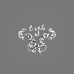
| Projections & slices | Image control
Images are generated by Spider. | ||||||||||||||||||||||||||||||||||||||||||||||||||||||||||||||||||||
| Voxel size | X=Y=Z: 1.62 Å | ||||||||||||||||||||||||||||||||||||||||||||||||||||||||||||||||||||
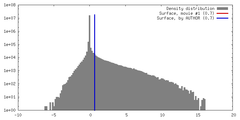
| Density |
| ||||||||||||||||||||||||||||||||||||||||||||||||||||||||||||||||||||
| Symmetry | Space group: 1 | ||||||||||||||||||||||||||||||||||||||||||||||||||||||||||||||||||||
| Details | EMDB XML:
CCP4 map header:
| ||||||||||||||||||||||||||||||||||||||||||||||||||||||||||||||||||||
 Z (Sec.)
 Y (Row.)
 X (Col.)


-Supplemental data
- Sample components
-Entire : Inositol 1,4,5-trisphosphate receptor, type 1
| Entire | Name: Inositol 1,4,5-trisphosphate receptor, type 1 |
|---|---|
| Components |
|
-Supramolecule #1000: Inositol 1,4,5-trisphosphate receptor, type 1
| Supramolecule | Name: Inositol 1,4,5-trisphosphate receptor, type 1 / type: sample / ID: 1000 Details: The sample was purified from rat cerebellum after solubilization with detergent; only freshly purified protein was used for cryo-EM visualization. Oligomeric state: tetramer / Number unique components: 1 |
|---|---|
| Molecular weight | Theoretical: 1.3 MDa |
-Macromolecule #1: Inositol 1,4,5-trisphosphate receptor
| Macromolecule | Name: Inositol 1,4,5-trisphosphate receptor / type: protein_or_peptide / ID: 1 / Name.synonym: Calcium release channel / Details: detergent-solubilized protein / Number of copies: 4 / Oligomeric state: tetramer / Recombinant expression: No / Database: NCBI |
|---|---|
| Source (natural) | Organism: |
| Molecular weight | Experimental: 300 KDa / Theoretical: 330 KDa |
| Sequence | UniProtKB: Inositol 1,4,5-trisphosphate-gated calcium channel ITPR1 |
-Experimental details
-Structure determination
| Method | cryo EM |
|---|---|
 Processing Processing | single particle reconstruction |
| Aggregation state | particle |
-Sample preparation
| Concentration | 0.4 mg/mL |
|---|---|
| Buffer | pH: 7.4 Details: 50 mM Tris-HCl, pH 7.4, 0.4% CHAPS, 150 mM NaCl, 1 mM DTT, 1 mM EGTA, 1 mM EDTA |
| Grid | Details: 400 mesh copper grids with thin carbon support |
| Vitrification | Cryogen name: ETHANE / Chamber humidity: 100 % / Chamber temperature: 120 K / Instrument: FEI VITROBOT MARK IV / Method: blot once for 3 seconds |
- Electron microscopy
| Microscope | FEI POLARA 300 |
|---|---|
| Temperature | Min: 95 K / Max: 102 K / Average: 100 K |
| Alignment procedure | Legacy - Astigmatism: Objective lens astigmatism was corrected at 100,000 times magnification. |
| Specialist optics | Energy filter - Name: FEI |
| Date | Jan 1, 2014 |
| Image recording | Category: CCD / Film or detector model: GATAN K2 (4k x 4k) / Digitization - Sampling interval: 0.81 µm / Number real images: 4160 / Average electron dose: 22 e/Å2 Details: Every image is the sum of 30 frames recorded with a direct electron detector. |
| Electron beam | Acceleration voltage: 300 kV / Electron source:  FIELD EMISSION GUN FIELD EMISSION GUN |
| Electron optics | Calibrated magnification: 30886 / Illumination mode: FLOOD BEAM / Imaging mode: BRIGHT FIELD / Cs: 2.0 mm / Nominal defocus max: 3.5 µm / Nominal defocus min: 0.6 µm / Nominal magnification: 23000 |
| Sample stage | Specimen holder model: GATAN LIQUID NITROGEN |
| Experimental equipment |  Model: Tecnai Polara / Image courtesy: FEI Company |
-Image processing
| Details | Particle selection: EMAN2.1, CTF correction: CTFFIND3, initial model: EMAN2.1, refinement: RELION 1.3 |
|---|---|
| CTF correction | Details: CTFFIND3 |
| Final reconstruction | Algorithm: OTHER / Resolution.type: BY AUTHOR / Resolution: 4.7 Å / Resolution method: OTHER / Software - Name: RELION_1.3, EMAN_2.1 / Details: The final map was generated from 96,106 particles. / Number images used: 96106 |
