 Movie
Movie Controller


+ Open data

- Basic information
| Entry | Database: PDB / ID: 5wfe | |||||||||
|---|---|---|---|---|---|---|---|---|---|---|
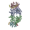
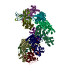
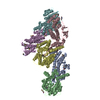
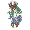
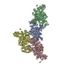
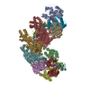
| Title | Cas1-Cas2-IHF-DNA holo-complex | |||||||||
 Components Components |
| |||||||||
 Keywords Keywords | DNA BINDING PROTEIN/DNA / CRISPR integration complex / DNA / Cas1-Cas2 / IHF / DNA BINDING PROTEIN / DNA BINDING PROTEIN-DNA complex | |||||||||
| Function / homology |  Function and homology information Function and homology informationCRISPR-cas system / positive regulation of metabolic process / crossover junction DNA endonuclease activity / 5'-flap endonuclease activity / maintenance of CRISPR repeat elements / structural constituent of chromatin / regulation of translation / chromosome / endonuclease activity / DNA recombination ...CRISPR-cas system / positive regulation of metabolic process / crossover junction DNA endonuclease activity / 5'-flap endonuclease activity / maintenance of CRISPR repeat elements / structural constituent of chromatin / regulation of translation / chromosome / endonuclease activity / DNA recombination / defense response to virus / Hydrolases; Acting on ester bonds / DNA repair / hydrolase activity / DNA damage response / regulation of DNA-templated transcription / DNA-templated transcription / protein homodimerization activity / DNA binding / metal ion binding / identical protein binding / cytoplasm / cytosol Similarity search - Function | |||||||||
| Biological species |  synthetic construct (others) | |||||||||
| Method | ELECTRON MICROSCOPY / single particle reconstruction / cryo EM / Resolution: 3.64 Å | |||||||||
 Authors Authors | Wright, A.V. / Liu, J.J. / Nogales, E. / Doudna, J.A. | |||||||||
| Funding support |  United States, 2items United States, 2items
| |||||||||
 Citation Citation | Journal: Science / Year: 2017 Title: Structures of the CRISPR genome integration complex. Authors: Addison V Wright / Jun-Jie Liu / Gavin J Knott / Kevin W Doxzen / Eva Nogales / Jennifer A Doudna / Abstract: CRISPR-Cas systems depend on the Cas1-Cas2 integrase to capture and integrate short foreign DNA fragments into the CRISPR locus, enabling adaptation to new viruses. We present crystal structures of ...CRISPR-Cas systems depend on the Cas1-Cas2 integrase to capture and integrate short foreign DNA fragments into the CRISPR locus, enabling adaptation to new viruses. We present crystal structures of Cas1-Cas2 bound to both donor and target DNA in intermediate and product integration complexes, as well as a cryo-electron microscopy structure of the full CRISPR locus integration complex, including the accessory protein IHF (integration host factor). The structures show unexpectedly that indirect sequence recognition dictates integration site selection by favoring deformation of the repeat and the flanking sequences. IHF binding bends the DNA sharply, bringing an upstream recognition motif into contact with Cas1 to increase both the specificity and efficiency of integration. These results explain how the Cas1-Cas2 CRISPR integrase recognizes a sequence-dependent DNA structure to ensure site-selective CRISPR array expansion during the initial step of bacterial adaptive immunity. | |||||||||
| History |
|
- Structure visualization
| Movie |
Movie viewer |
|---|---|
| Structure viewer | Molecule: MolmilJmol/JSmol |
- Downloads & links
-Download
| PDBx/mmCIF format | 5wfe.cif.gz | 391.6 KB | Display | PDBx/mmCIF format |
|---|---|---|---|---|
| PDB format | pdb5wfe.ent.gz | 306.9 KB | Display | PDB format |
| PDBx/mmJSON format | 5wfe.json.gz | Tree view | PDBx/mmJSON format | |
| Others |  Other downloads Other downloads |
-Validation report
| Arichive directory | https://data.pdbj.org/pub/pdb/validation_reports/wf/5wfeftp://data.pdbj.org/pub/pdb/validation_reports/wf/5wfe | HTTPS FTP |
|---|
-Related structure data
| Related structure data | 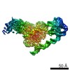 8827MC 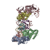 5vvjC 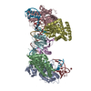 5vvkC 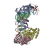 5vvlC M: map data used to model this data C: citing same article ( |
|---|---|
| Similar structure data |


-Links
 PDBj
PDBj


- Assembly
| Deposited unit | 
|
|---|---|
| 1 |
|
-Components
-CRISPR-associated ... , 2 types, 6 molecules ABCDEF
| #1: Protein | Mass: 33235.418 Da / Num. of mol.: 4 Source method: isolated from a genetically manipulated source Details: Cas1 / Source: (gene. exp.) References: UniProt: Q46896, Hydrolases; Acting on ester bonds #2: Protein | Mass: 11553.270 Da / Num. of mol.: 2 Source method: isolated from a genetically manipulated source Source: (gene. exp.) References: UniProt: P45956, Hydrolases; Acting on ester bonds |
|---|
-DNA chain , 4 types, 4 molecules GHIJ
| #3: DNA chain | Mass: 8680.646 Da / Num. of mol.: 1 / Source method: obtained synthetically / Source: (synth.) synthetic construct (others) |
|---|---|
| #4: DNA chain | Mass: 18745.965 Da / Num. of mol.: 1 / Source method: obtained synthetically / Source: (synth.) synthetic construct (others) |
| #5: DNA chain | Mass: 29101.656 Da / Num. of mol.: 1 / Source method: obtained synthetically / Source: (synth.) synthetic construct (others) |
| #6: DNA chain | Mass: 19286.447 Da / Num. of mol.: 1 / Source method: obtained synthetically / Source: (synth.) synthetic construct (others) |
-Integration host factor subunit ... , 2 types, 2 molecules KL
| #7: Protein | Mass: 11373.952 Da / Num. of mol.: 1 Source method: isolated from a genetically manipulated source Source: (gene. exp.) |
|---|---|
| #8: Protein | Mass: 10671.178 Da / Num. of mol.: 1 Source method: isolated from a genetically manipulated source Source: (gene. exp.) |
-Experimental details
-Experiment
| Experiment | Method: ELECTRON MICROSCOPY |
|---|---|
| EM experiment | Aggregation state: PARTICLE / 3D reconstruction method: single particle reconstruction |
- Sample preparation
| Component | Name: Cas1-Cas2-IHF-DNA holo complex / Type: COMPLEX / Entity ID: all / Source: RECOMBINANT |
|---|---|
| Molecular weight | Experimental value: NO |
| Source (natural) | Organism: |
| Source (recombinant) | Organism: |
| Buffer solution | pH: 7.5 Details: 20 mM HEPES, pH 7.5, 150 mM KCl, 5 mM EDTA, 1 mM DTT, and 0.1% glycerol |
| Specimen | Conc.: 0.3 mg/ml / Embedding applied: NO / Shadowing applied: NO / Staining applied: NO / Vitrification applied: YES / Details: 1uM Cas1-Cas2-DNA-IHF complexes |
| Specimen support | Grid material: COPPER / Grid mesh size: 400 divisions/in. / Grid type: C-flat |
| Vitrification | Instrument: FEI VITROBOT MARK IV / Cryogen name: ETHANE / Humidity: 100 % / Chamber temperature: 281.15 K |
- Electron microscopy imaging
| Experimental equipment |  Model: Titan Krios / Image courtesy: FEI Company |
|---|---|
| Microscopy | Model: FEI TITAN KRIOS |
| Electron gun | Electron source:  FIELD EMISSION GUN / Accelerating voltage: 300 kV / Illumination mode: FLOOD BEAM FIELD EMISSION GUN / Accelerating voltage: 300 kV / Illumination mode: FLOOD BEAM |
| Electron lens | Mode: BRIGHT FIELD / Cs: 2.6 mm / Alignment procedure: COMA FREE |
| Specimen holder | Cryogen: NITROGEN / Specimen holder model: FEI TITAN KRIOS AUTOGRID HOLDER |
| Image recording | Average exposure time: 6 sec. / Electron dose: 1.5 e/Å2 / Detector mode: SUPER-RESOLUTION / Film or detector model: GATAN K2 SUMMIT (4k x 4k) / Num. of grids imaged: 1 / Num. of real images: 3000 |
| Image scans | Movie frames/image: 30 / Used frames/image: 3-30 |
- Processing
| Software | Name: PHENIX / Version: 1.11.1_2575: / Classification: refinement | ||||||||||||||||||||||||||||||||||||||||
|---|---|---|---|---|---|---|---|---|---|---|---|---|---|---|---|---|---|---|---|---|---|---|---|---|---|---|---|---|---|---|---|---|---|---|---|---|---|---|---|---|---|
| EM software |
| ||||||||||||||||||||||||||||||||||||||||
| CTF correction | Type: PHASE FLIPPING AND AMPLITUDE CORRECTION | ||||||||||||||||||||||||||||||||||||||||
| Particle selection | Num. of particles selected: 650000 | ||||||||||||||||||||||||||||||||||||||||
| Symmetry | Point symmetry: C1 (asymmetric) | ||||||||||||||||||||||||||||||||||||||||
| 3D reconstruction | Resolution: 3.64 Å / Resolution method: FSC 0.143 CUT-OFF / Num. of particles: 86000 / Symmetry type: POINT | ||||||||||||||||||||||||||||||||||||||||
| Atomic model building |
| ||||||||||||||||||||||||||||||||||||||||
| Refine LS restraints |
|
