 Movie
Movie Controller


+ Open data

- Basic information
| Entry | Database: PDB / ID: 5vy5 | ||||||
|---|---|---|---|---|---|---|---|
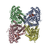
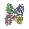
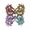
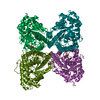
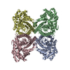
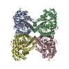
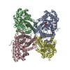
| Title | Rabbit muscle aldolase using 200keV | ||||||
 Components Components | Fructose-bisphosphate aldolase A | ||||||
 Keywords Keywords | LYASE / glycolytic enzyme | ||||||
| Function / homology |  Function and homology information Function and homology informationnegative regulation of Arp2/3 complex-mediated actin nucleation / fructose-bisphosphate aldolase / fructose-bisphosphate aldolase activity / M band / I band / glycolytic process / protein homotetramerization / positive regulation of cell migration Similarity search - Function | ||||||
| Biological species |  | ||||||
| Method | ELECTRON MICROSCOPY / single particle reconstruction / cryo EM / Resolution: 2.6 Å | ||||||
 Authors Authors | Herzik Jr., M.A. / Wu, M. / Lander, G.C. | ||||||
| Funding support |  United States, 1items United States, 1items
| ||||||
 Citation Citation | Journal: Nat Methods / Year: 2017 Title: Achieving better-than-3-Å resolution by single-particle cryo-EM at 200 keV. Authors: Mark A Herzik / Mengyu Wu / Gabriel C Lander / Abstract: Nearly all single-particle cryo-EM structures resolved to better than 4-Å resolution have been determined using 300-keV transmission electron microscopes (TEMs). We demonstrate that it is possible ...Nearly all single-particle cryo-EM structures resolved to better than 4-Å resolution have been determined using 300-keV transmission electron microscopes (TEMs). We demonstrate that it is possible to obtain reconstructions of macromolecular complexes of different sizes to better than 3-Å resolution using a 200-keV TEM. These structures are of sufficient quality to unambiguously assign amino acid rotameric conformations and identify ordered water molecules. | ||||||
| History |
|
- Structure visualization
| Movie |
Movie viewer |
|---|---|
| Structure viewer | Molecule: MolmilJmol/JSmol |
- Downloads & links
-Download
| PDBx/mmCIF format | 5vy5.cif.gz | 2.1 MB | Display | PDBx/mmCIF format |
|---|---|---|---|---|
| PDB format | pdb5vy5.ent.gz | 1.8 MB | Display | PDB format |
| PDBx/mmJSON format | 5vy5.json.gz | Tree view | PDBx/mmJSON format | |
| Others |  Other downloads Other downloads |
-Validation report
| Arichive directory | https://data.pdbj.org/pub/pdb/validation_reports/vy/5vy5ftp://data.pdbj.org/pub/pdb/validation_reports/vy/5vy5 | HTTPS FTP |
|---|
-Related structure data
| Related structure data | 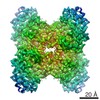 8743MC 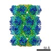 8741C 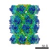 8742C 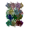 5vy3C  5vy4C M: map data used to model this data C: citing same article ( |
|---|---|
| Similar structure data | |
| EM raw data | EMPIAR-10181 (Title: Rabbit muscle aldolase movies obtained using Talos Arctica operating at 200 kV equipped with a K2 Data size: 1.5 TB Data #1: Raw, unaligned movie stacks of rabbit muscle aldolase acquired on a Talos Arctica using a K2 direct electron detector [micrographs - multiframe]) |


-Links
 PDBj
PDBj
- Assembly
| Deposited unit | 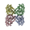
|
|---|---|
| 1 |
|
| Number of models | 10 |
-Components
| #1: Protein | Mass: 39263.672 Da / Num. of mol.: 4 Source method: isolated from a genetically manipulated source Source: (gene. exp.)  |
|---|
-Experimental details
-Experiment
| Experiment | Method: ELECTRON MICROSCOPY |
|---|---|
| EM experiment | Aggregation state: PARTICLE / 3D reconstruction method: single particle reconstruction |
- Sample preparation
| Component | Name: Rabbit Muscle Aldolase / Type: COMPLEX Details: Rabbit Muscle Aldolase (reconstituted from lyophilized form) Entity ID: all / Source: RECOMBINANT | |||||||||||||||
|---|---|---|---|---|---|---|---|---|---|---|---|---|---|---|---|---|
| Molecular weight | Value: 0.15 MDa / Experimental value: NO | |||||||||||||||
| Source (natural) | Organism: | |||||||||||||||
| Source (recombinant) | Organism: | |||||||||||||||
| Buffer solution | pH: 7.5 | |||||||||||||||
| Buffer component |
| |||||||||||||||
| Specimen | Conc.: 1.6 mg/ml / Embedding applied: NO / Shadowing applied: NO / Staining applied: NO / Vitrification applied: YES Details: Specimen was prepared from lyophilized rabbit muscle aldolase (Sigma Aldrich) | |||||||||||||||
| Specimen support | Details: 15 Watts / Grid material: GOLD / Grid mesh size: 300 divisions/in. / Grid type: Quantifoil, UltrAuFoil, R1.2/1.3 | |||||||||||||||
| Vitrification | Instrument: HOMEMADE PLUNGER / Cryogen name: ETHANE / Humidity: 100 % / Chamber temperature: 277.15 K Details: 3 uL of sample/grid was manually blotted for 4 seconds prior to immediate plunge-freezing in liquid nitrogen-cooled ethane. |
- Electron microscopy imaging
| Experimental equipment |  Model: Talos Arctica / Image courtesy: FEI Company |
|---|---|
| Microscopy | Model: FEI TALOS ARCTICA |
| Electron gun | Electron source:  FIELD EMISSION GUN / Accelerating voltage: 200 kV / Illumination mode: FLOOD BEAM FIELD EMISSION GUN / Accelerating voltage: 200 kV / Illumination mode: FLOOD BEAM |
| Electron lens | Mode: BRIGHT FIELD / Nominal magnification: 45000 X / Nominal defocus max: 2700 nm / Nominal defocus min: 600 nm / Cs: 2.7 mm / C2 aperture diameter: 70 µm / Alignment procedure: COMA FREE |
| Specimen holder | Cryogen: NITROGEN |
| Image recording | Average exposure time: 11 sec. / Electron dose: 68 e/Å2 / Detector mode: SUPER-RESOLUTION / Film or detector model: GATAN K2 SUMMIT (4k x 4k) / Num. of grids imaged: 1 / Num. of real images: 810 Details: Images collected using stage position navigation to target exposure. |
| Image scans | Sampling size: 5 µm / Width: 7420 / Height: 7676 / Movie frames/image: 44 / Used frames/image: 1-44 |
- Processing
| Software | Name: PHENIX / Version: 1.10.1_2155: / Classification: refinement | |||||||||||||||||||||||||||||||||||||||||||||||||||||||
|---|---|---|---|---|---|---|---|---|---|---|---|---|---|---|---|---|---|---|---|---|---|---|---|---|---|---|---|---|---|---|---|---|---|---|---|---|---|---|---|---|---|---|---|---|---|---|---|---|---|---|---|---|---|---|---|---|
| EM software |
| |||||||||||||||||||||||||||||||||||||||||||||||||||||||
| Image processing | Details: Movies were collected in super-resolution mode and Fourier-binned by two prior to motion correction and dose weighting using MotionCor2 program. | |||||||||||||||||||||||||||||||||||||||||||||||||||||||
| CTF correction | Type: PHASE FLIPPING AND AMPLITUDE CORRECTION | |||||||||||||||||||||||||||||||||||||||||||||||||||||||
| Particle selection | Num. of particles selected: 1009341 Details: Template-based particle picking was performed using templates generated from reference-free 2D classification of an initial set of automated particle picks. | |||||||||||||||||||||||||||||||||||||||||||||||||||||||
| Symmetry | Point symmetry: D2 (2x2 fold dihedral) | |||||||||||||||||||||||||||||||||||||||||||||||||||||||
| 3D reconstruction | Resolution: 2.6 Å / Resolution method: FSC 0.143 CUT-OFF / Num. of particles: 83910 / Algorithm: BACK PROJECTION / Symmetry type: POINT | |||||||||||||||||||||||||||||||||||||||||||||||||||||||
| Atomic model building | B value: 35 / Protocol: OTHER / Space: REAL / Target criteria: Maximum Likelihood Details: Starting model was generated by stripping PDB entry 6ALD of all ligands and alternate conformations, then refining into the EM density using imposed symmetry while adjusting ...Details: Starting model was generated by stripping PDB entry 6ALD of all ligands and alternate conformations, then refining into the EM density using imposed symmetry while adjusting weighting/scoring according to estimated map resolution. The top 10 generated models (ranked based on quality metrics) were real-space refined using Phenix software. | |||||||||||||||||||||||||||||||||||||||||||||||||||||||
| Refine LS restraints |
|
