Movie
Movie Controller


+ Open data

- Basic information
| Entry | Database: PDB / ID: 5jxl | ||||||
|---|---|---|---|---|---|---|---|
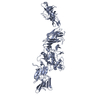
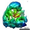
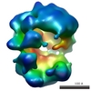
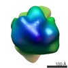
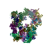
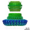
| Title | Cryo-EM structure of the flagellar hook of Campylobacter jejuni | ||||||
 Components Components | flagellar hook protein FlgE | ||||||
 Keywords Keywords | MOTOR PROTEIN / Campylobacter jejuni / helical assembly of FlgE / flagellar hook | ||||||
| Function / homology |  Function and homology information Function and homology informationbacterial-type flagellum organization / bacterial-type flagellum basal body / bacterial-type flagellum-dependent swarming motility Similarity search - Function | ||||||
| Biological species |  Campylobacter jejuni subsp. jejuni 81116 (Campylobacter) Campylobacter jejuni subsp. jejuni 81116 (Campylobacter) | ||||||
| Method | ELECTRON MICROSCOPY / helical reconstruction / cryo EM / Resolution: 3.5 Å | ||||||
 Authors Authors | Matsunami, H. / Wolf, M. / Samatey, F.A. | ||||||
| Funding support |  Japan, 1items Japan, 1items
| ||||||
 Citation Citation | Journal: Nat Commun / Year: 2016 Title: Complete structure of the bacterial flagellar hook reveals extensive set of stabilizing interactions. Authors: Hideyuki Matsunami / Clive S Barker / Young-Ho Yoon / Matthias Wolf / Fadel A Samatey / Abstract: The bacterial flagellar hook is a tubular helical structure made by the polymerization of multiple copies of a protein, FlgE. Here we report the structure of the hook from Campylobacter jejuni by ...The bacterial flagellar hook is a tubular helical structure made by the polymerization of multiple copies of a protein, FlgE. Here we report the structure of the hook from Campylobacter jejuni by cryo-electron microscopy at a resolution of 3.5 Å. On the basis of this structure, we show that the hook is stabilized by intricate inter-molecular interactions between FlgE molecules. Extra domains in FlgE, found only in Campylobacter and in related bacteria, bring more stability and robustness to the hook. Functional experiments suggest that Campylobacter requires an unusually strong hook to swim without its flagella being torn off. This structure reveals details of the quaternary organization of the hook that consists of 11 protofilaments. Previous study of the flagellar filament of Campylobacter by electron microscopy showed its quaternary structure made of seven protofilaments. Therefore, this study puts in evidence the difference between the quaternary structures of a bacterial filament and its hook. | ||||||
| History |
|
- Structure visualization
| Movie |
Movie viewer |
|---|---|
| Structure viewer | Molecule: MolmilJmol/JSmol |
- Downloads & links
-Download
| PDBx/mmCIF format | 5jxl.cif.gz | 148.1 KB | Display | PDBx/mmCIF format |
|---|---|---|---|---|
| PDB format | pdb5jxl.ent.gz | 112.1 KB | Display | PDB format |
| PDBx/mmJSON format | 5jxl.json.gz | Tree view | PDBx/mmJSON format | |
| Others |  Other downloads Other downloads |
-Validation report
| Arichive directory | https://data.pdbj.org/pub/pdb/validation_reports/jx/5jxlftp://data.pdbj.org/pub/pdb/validation_reports/jx/5jxl | HTTPS FTP |
|---|
-Related structure data
| Related structure data | 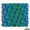 8179MC M: map data used to model this data C: citing same article ( |
|---|---|
| Similar structure data |

-Links
 PDBj
PDBj
- Assembly
| Deposited unit | 
|
|---|---|
| 1 | x 11
|
| 2 |
|
| 3 | 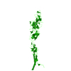
|
| Symmetry | Helical symmetry: (Circular symmetry: 1 / Dyad axis: no / N subunits divisor: 1 / Num. of operations: 11 / Rise per n subunits: 4.185 Å / Rotation per n subunits: 64.34 °) |
-Components
| #1: Protein | Mass: 90449.750 Da / Num. of mol.: 1 / Source method: isolated from a natural source Source: (natural) Campylobacter jejuni subsp. jejuni 81116 (Campylobacter)Strain: 81116 / References: UniProt: A0A1L1QK18*PLUS |
|---|
-Experimental details
-Experiment
| Experiment | Method: ELECTRON MICROSCOPY |
|---|---|
| EM experiment | Aggregation state: FILAMENT / 3D reconstruction method: helical reconstruction |
- Sample preparation
| Component | Name: Campylobacter Hook / Type: ORGANELLE OR CELLULAR COMPONENT / Entity ID: all / Source: NATURAL |
|---|---|
| Molecular weight | Experimental value: NO |
| Source (natural) | Organism:  Campylobacter jejuni (Campylobacter) Campylobacter jejuni (Campylobacter) |
| Buffer solution | pH: 8 / Details: EDTA, Triton X-100 |
| Buffer component | Conc.: 10 mM / Name: Tris Hydrochloride / Formula: Tris-HCl |
| Specimen | Embedding applied: NO / Shadowing applied: NO / Staining applied: NO / Vitrification applied: YES |
| Specimen support | Grid material: COPPER / Grid mesh size: 400 divisions/in. / Grid type: C-flat-1.2/1.3 |
| Vitrification | Instrument: GATAN CRYOPLUNGE 3 / Cryogen name: ETHANE / Humidity: 90 % / Chamber temperature: 280 K Details: blotting from both side, Whatman #40 filter paper, 25 seconds blot time |
- Electron microscopy imaging
| Experimental equipment |  Model: Titan Krios / Image courtesy: FEI Company |
|---|---|
| Microscopy | Model: FEI TITAN KRIOS / Details: parallel illumination |
| Electron gun | Electron source:  FIELD EMISSION GUN / Accelerating voltage: 300 kV / Illumination mode: FLOOD BEAM FIELD EMISSION GUN / Accelerating voltage: 300 kV / Illumination mode: FLOOD BEAM |
| Electron lens | Mode: BRIGHT FIELD / Nominal magnification: 79000 X / Calibrated magnification: 105000 X / Nominal defocus max: 2500 nm / Nominal defocus min: 500 nm / Calibrated defocus min: 500 nm / Calibrated defocus max: 2500 nm / Cs: 2.7 mm / C2 aperture diameter: 100 µm / Alignment procedure: COMA FREE |
| Specimen holder | Cryogen: NITROGEN / Specimen holder model: FEI TITAN KRIOS AUTOGRID HOLDER / Temperature (max): 70 K / Temperature (min): 63 K |
| Image recording | Average exposure time: 1.5 sec. / Electron dose: 2.4 e/Å2 / Detector mode: INTEGRATING / Film or detector model: FEI FALCON II (4k x 4k) / Num. of grids imaged: 1 / Num. of real images: 504 |
| Image scans | Sampling size: 14 µm / Width: 4096 / Height: 4096 / Movie frames/image: 18 / Used frames/image: 1-5 |
- Processing
| EM software |
| ||||||||||||||||||||||||||||||||||||||||||||||||||||||||||||
|---|---|---|---|---|---|---|---|---|---|---|---|---|---|---|---|---|---|---|---|---|---|---|---|---|---|---|---|---|---|---|---|---|---|---|---|---|---|---|---|---|---|---|---|---|---|---|---|---|---|---|---|---|---|---|---|---|---|---|---|---|---|
| Image processing | Details: first 5 images were aligned for drift correction and summed. Summed images were normalizaed. | ||||||||||||||||||||||||||||||||||||||||||||||||||||||||||||
| CTF correction | Details: SPRING v0.83 / Type: PHASE FLIPPING ONLY | ||||||||||||||||||||||||||||||||||||||||||||||||||||||||||||
| Helical symmerty | Angular rotation/subunit: 64.34 ° / Axial rise/subunit: 4.185 Å / Axial symmetry: C1 | ||||||||||||||||||||||||||||||||||||||||||||||||||||||||||||
| Particle selection | Num. of particles selected: 70477 | ||||||||||||||||||||||||||||||||||||||||||||||||||||||||||||
| 3D reconstruction | Resolution: 3.5 Å / Resolution method: FSC 0.143 CUT-OFF / Num. of particles: 49884 / Algorithm: FOURIER SPACE / Symmetry type: HELICAL | ||||||||||||||||||||||||||||||||||||||||||||||||||||||||||||
| Atomic model building | B value: 120 / Protocol: BACKBONE TRACE / Space: REAL / Target criteria: CC / Details: side chains included, default parameters | ||||||||||||||||||||||||||||||||||||||||||||||||||||||||||||
| Atomic model building | PDB-ID: 5AZ4 Pdb chain-ID: A / Accession code: 5AZ4 / Source name: PDB / Type: experimental model |