 Movie
Movie Controller

[English] 日本語
 Yorodumi
- PDB-5mju: Structure of the thermostabilized EAAT1 cryst mutant in complex w... -
+ Open data

- Basic information
| Entry | Database: PDB / ID: 5mju | ||||||
|---|---|---|---|---|---|---|---|
| Title | Structure of the thermostabilized EAAT1 cryst mutant in complex with the competititve inhibitor TFB-TBOA and the allosteric inhibitor UCPH101 | ||||||
 Components Components | Excitatory amino acid transporter 1,Neutral amino acid transporter B(0),Excitatory amino acid transporter 1 | ||||||
 Keywords Keywords | TRANSPORT PROTEIN / excitatory aminoacid transporter 1 / human glutamate transporter / TFB-TBOA / UCPH-101 | ||||||
| Function / homology |  Function and homology information Function and homology informationDefective SLC1A3 causes episodic ataxia 6 (EA6) / cranial nerve development / Astrocytic Glutamate-Glutamine Uptake And Metabolism / L-glutamine secretion / GABA biosynthetic process / neurotransmitter uptake / cell morphogenesis involved in neuron differentiation / L-glutamine import across plasma membrane / L-glutamine transmembrane transporter activity / L-serine transmembrane transporter activity ...Defective SLC1A3 causes episodic ataxia 6 (EA6) / cranial nerve development / Astrocytic Glutamate-Glutamine Uptake And Metabolism / L-glutamine secretion / GABA biosynthetic process / neurotransmitter uptake / cell morphogenesis involved in neuron differentiation / L-glutamine import across plasma membrane / L-glutamine transmembrane transporter activity / L-serine transmembrane transporter activity / L-glutamine transport / high-affinity L-glutamate transmembrane transporter activity / glutamate:sodium symporter activity / L-glutamate import / : / membrane protein complex / L-glutamate transmembrane transporter activity / L-glutamate transmembrane transport / ligand-gated channel activity / L-aspartate transmembrane transporter activity / neutral amino acid transport / Glutamate Neurotransmitter Release Cycle / L-aspartate import across plasma membrane / D-aspartate import across plasma membrane / neutral L-amino acid transmembrane transporter activity / auditory behavior / L-glutamate import across plasma membrane / intracellular sodium ion homeostasis / transepithelial transport / symporter activity / Amino acid transport across the plasma membrane / amino acid transmembrane transporter activity / cellular response to cocaine / antiporter activity / neuromuscular process controlling balance / glutamate binding / RHOJ GTPase cycle / protein homotrimerization / RHOQ GTPase cycle / neurotransmitter transport / amino acid transport / RHOH GTPase cycle / RAC3 GTPase cycle / response to light stimulus / positive regulation of synaptic transmission / transport across blood-brain barrier / RAC1 GTPase cycle / monoatomic ion transport / potassium ion transmembrane transport / basal plasma membrane / chloride transmembrane transport / erythrocyte differentiation / sensory perception of sound / response to wounding / centriolar satellite / melanosome / virus receptor activity / signaling receptor activity / cytoplasmic vesicle / chemical synaptic transmission / neuron projection / ciliary basal body / response to xenobiotic stimulus / response to antibiotic / neuronal cell body / synapse / perinuclear region of cytoplasm / cell surface / extracellular exosome / membrane / metal ion binding / plasma membrane Similarity search - Function | ||||||
| Biological species |  Homo sapiens (human) Homo sapiens (human) | ||||||
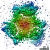
| Method |  X-RAY DIFFRACTION / SYNCHROTRON / MOLECULAR REPLACEMENT / Resolution: 3.71 Å X-RAY DIFFRACTION / SYNCHROTRON / MOLECULAR REPLACEMENT / Resolution: 3.71 Å | ||||||
 Authors Authors | Canul-Tec, J. / Assal, R. / Legrand, P. / Reyes, N. | ||||||
| Funding support |  France, 1items France, 1items
| ||||||
 Citation Citation | Journal: Nature / Year: 2017 Title: Structure and allosteric inhibition of excitatory amino acid transporter 1. Authors: Canul-Tec, J.C. / Assal, R. / Cirri, E. / Legrand, P. / Brier, S. / Chamot-Rooke, J. / Reyes, N. | ||||||
| History |
|
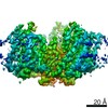
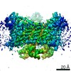
- Structure visualization
| Structure viewer | Molecule: MolmilJmol/JSmol |
|---|
- Downloads & links
-Download
| PDBx/mmCIF format | 5mju.cif.gz | 173.5 KB | Display | PDBx/mmCIF format |
|---|---|---|---|---|
| PDB format | pdb5mju.ent.gz | 138.2 KB | Display | PDB format |
| PDBx/mmJSON format | 5mju.json.gz | Tree view | PDBx/mmJSON format | |
| Others |  Other downloads Other downloads |
-Validation report
| Arichive directory | https://data.pdbj.org/pub/pdb/validation_reports/mj/5mjuftp://data.pdbj.org/pub/pdb/validation_reports/mj/5mju | HTTPS FTP |
|---|
-Related structure data
| Related structure data |  5llmSC 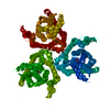 5lluC  5lm4C S: Starting model for refinement C: citing same article ( |
|---|---|
| Similar structure data |


-Links
 PDBj
PDBj


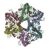
- Assembly
| Deposited unit | 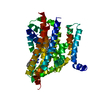
| ||||||||
|---|---|---|---|---|---|---|---|---|---|
| 1 | 
| ||||||||
| Unit cell |
|
-Components
| #1: Protein | Mass: 56512.379 Da / Num. of mol.: 1 Source method: isolated from a genetically manipulated source Source: (gene. exp.) Homo sapiens (human)Gene: SLC1A3, EAAT1, GLAST, GLAST1, SLC1A5, ASCT2, M7V1, RDR, RDRC Plasmid: pcDNA3 / Cell (production host): Epithelial / Cell line (production host): HEK-293F / Organ (production host): Kidney / Production host: Homo sapiens (human) / Tissue (production host): Embryonic kidney / References: UniProt: P43003, UniProt: Q15758 |
|---|---|
| #2: Chemical | ChemComp-6Z6 / 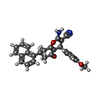  Mass: 422.475 Da / Num. of mol.: 1 / Source method: obtained synthetically / Formula: C27H22N2O3 Mass: 422.475 Da / Num. of mol.: 1 / Source method: obtained synthetically / Formula: C27H22N2O3 |
| #3: Chemical | ChemComp-7O9 / (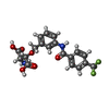  Mass: 426.343 Da / Num. of mol.: 1 / Source method: obtained synthetically / Formula: C19H17F3N2O6 Mass: 426.343 Da / Num. of mol.: 1 / Source method: obtained synthetically / Formula: C19H17F3N2O6 |
-Experimental details
-Experiment
| Experiment | Method: X-RAY DIFFRACTION / Number of used crystals: 1 |
|---|
- Sample preparation
| Crystal | Density Matthews: 3.59 Å3/Da / Density % sol: 64.8 % |
|---|---|
| Crystal grow | Temperature: 277 K / Method: vapor diffusion, hanging drop / pH: 8.2 Details: 32% PEG 400, 100 mM Tris pH 8.2, 50 mM Calcium chloride, 50 mM Barium chloride PH range: 8.0 - 8.4 |
-Data collection
| Diffraction | Mean temperature: 100 K |
|---|---|
| Diffraction source | Source: SYNCHROTRON / Site: ESRF / Beamline: ID30B / Wavelength: 0.9772 Å |
| Detector | Type: DECTRIS PILATUS 6M-F / Detector: PIXEL / Date: Oct 28, 2016 |
| Radiation | Monochromator: channel cut monocromator / Protocol: SINGLE WAVELENGTH / Monochromatic (M) / Laue (L): M / Scattering type: x-ray |
| Radiation wavelength | Wavelength: 0.9772 Å / Relative weight: 1 |
| Reflection | Resolution: 3.71→46.31 Å / Num. obs: 8570 / % possible obs: 99.9 % / Redundancy: 16.6 % / Biso Wilson estimate: 109.76 Å2 / CC1/2: 0.999 / Rmerge(I) obs: 0.14 / Rsym value: 0.051 / Net I/σ(I): 12.1 |
| Reflection shell | Resolution: 3.71→3.81 Å / Redundancy: 17.7 % / Rmerge(I) obs: 3.7 / Mean I/σ(I) obs: 0.9 / CC1/2: 0.373 / % possible all: 100 |
- Processing
| Software |
| ||||||||||||||||||||||||||||||||||||||||||||||||||||||||||||||||||||||||||||||||||||||||||||||||||||||||||||||||||
|---|---|---|---|---|---|---|---|---|---|---|---|---|---|---|---|---|---|---|---|---|---|---|---|---|---|---|---|---|---|---|---|---|---|---|---|---|---|---|---|---|---|---|---|---|---|---|---|---|---|---|---|---|---|---|---|---|---|---|---|---|---|---|---|---|---|---|---|---|---|---|---|---|---|---|---|---|---|---|---|---|---|---|---|---|---|---|---|---|---|---|---|---|---|---|---|---|---|---|---|---|---|---|---|---|---|---|---|---|---|---|---|---|---|---|---|
| Refinement | Method to determine structure: MOLECULAR REPLACEMENT Starting model: 5LLM Resolution: 3.71→25 Å / Cor.coef. Fo:Fc: 0.8455 / Cor.coef. Fo:Fc free: 0.7923 / Cross valid method: THROUGHOUT / σ(F): 0 / SU Rfree Blow DPI: 0.65
| ||||||||||||||||||||||||||||||||||||||||||||||||||||||||||||||||||||||||||||||||||||||||||||||||||||||||||||||||||
| Displacement parameters | Biso mean: 135.41 Å2
| ||||||||||||||||||||||||||||||||||||||||||||||||||||||||||||||||||||||||||||||||||||||||||||||||||||||||||||||||||
| Refine analyze | Luzzati coordinate error obs: 0.561 Å | ||||||||||||||||||||||||||||||||||||||||||||||||||||||||||||||||||||||||||||||||||||||||||||||||||||||||||||||||||
| Refinement step | Cycle: 1 / Resolution: 3.71→25 Å
| ||||||||||||||||||||||||||||||||||||||||||||||||||||||||||||||||||||||||||||||||||||||||||||||||||||||||||||||||||
| Refine LS restraints |
| ||||||||||||||||||||||||||||||||||||||||||||||||||||||||||||||||||||||||||||||||||||||||||||||||||||||||||||||||||
| LS refinement shell | Resolution: 3.71→4.15 Å / Total num. of bins used: 5
| ||||||||||||||||||||||||||||||||||||||||||||||||||||||||||||||||||||||||||||||||||||||||||||||||||||||||||||||||||
| Refinement TLS params. | Method: refined / Origin x: 53.9697 Å / Origin y: 155.7897 Å / Origin z: 1.539 Å
| ||||||||||||||||||||||||||||||||||||||||||||||||||||||||||||||||||||||||||||||||||||||||||||||||||||||||||||||||||
| Refinement TLS group | Selection details: { A|* } |
