[English] 日本語
Yorodumi
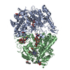
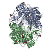
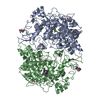
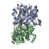
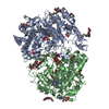
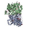
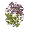
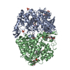
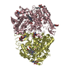
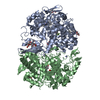
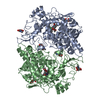
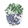
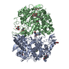
- PDB-5kir: The Structure of Vioxx Bound to Human COX-2 -

+
Open data


ID or keywords:

Loading...

-
Basic information

Entry
Database: PDB / ID: 5kir
TitleThe Structure of Vioxx Bound to Human COX-2
ComponentsProstaglandin G/H synthase 2
KeywordsOXIDOREDUCTASE / cyclooxyrgenase / Vioxx / Rofecoxib / COX
Function / homology
Function and homology information


Biosynthesis of EPA-derived SPMs / Biosynthesis of DPAn-3 SPMs / Biosynthesis of DHA-derived SPMs / Biosynthesis of electrophilic ω-3 PUFA oxo-derivatives / Synthesis of 15-eicosatetraenoic acid derivatives / cellular response to non-ionic osmotic stress / positive regulation of platelet-derived growth factor production / hair cycle / cellular response to homocysteine / positive regulation of fibroblast growth factor production ...Biosynthesis of EPA-derived SPMs / Biosynthesis of DPAn-3 SPMs / Biosynthesis of DHA-derived SPMs / Biosynthesis of electrophilic ω-3 PUFA oxo-derivatives / Synthesis of 15-eicosatetraenoic acid derivatives / cellular response to non-ionic osmotic stress / positive regulation of platelet-derived growth factor production / hair cycle / cellular response to homocysteine / positive regulation of fibroblast growth factor production / prostaglandin-endoperoxide synthase / prostaglandin-endoperoxide synthase activity / negative regulation of synaptic transmission, dopaminergic / cellular response to lead ion / response to nematode / positive regulation of transforming growth factor beta production / negative regulation of intrinsic apoptotic signaling pathway in response to osmotic stress / positive regulation of prostaglandin biosynthetic process / positive regulation of synaptic plasticity / regulation of neuroinflammatory response / Synthesis of Prostaglandins (PG) and Thromboxanes (TX) / Nicotinamide salvaging / response to fructose / cyclooxygenase pathway / positive regulation of smooth muscle contraction / response to fatty acid / positive regulation of fever generation / response to vitamin D / cellular response to fluid shear stress / prostaglandin secretion / oxidoreductase activity, acting on single donors with incorporation of molecular oxygen, incorporation of two atoms of oxygen / response to manganese ion / nuclear outer membrane / response to angiotensin / nuclear inner membrane / negative regulation of smooth muscle contraction / prostaglandin biosynthetic process / positive regulation of cell migration involved in sprouting angiogenesis / cellular response to ATP / maintenance of blood-brain barrier / bone mineralization / Interleukin-10 signaling / negative regulation of calcium ion transport / decidualization / negative regulation of cell cycle / positive regulation of vascular endothelial growth factor production / response to tumor necrosis factor / brown fat cell differentiation / response to glucocorticoid / positive regulation of vasoconstriction / positive regulation of brown fat cell differentiation / embryo implantation / positive regulation of synaptic transmission, glutamatergic / learning / positive regulation of smooth muscle cell proliferation / peroxidase activity / caveola / memory / regulation of blood pressure / cellular response to mechanical stimulus / positive regulation of protein import into nucleus / positive regulation of nitric oxide biosynthetic process / positive regulation of peptidyl-serine phosphorylation / response to estradiol / cellular response to heat / regulation of inflammatory response / cellular response to hypoxia / Interleukin-4 and Interleukin-13 signaling / angiogenesis / response to oxidative stress / response to lipopolysaccharide / neuron projection / positive regulation of apoptotic process / response to xenobiotic stimulus / negative regulation of cell population proliferation / endoplasmic reticulum lumen / heme binding / endoplasmic reticulum membrane / enzyme binding / endoplasmic reticulum / protein homodimerization activity / protein-containing complex / metal ion binding / cytoplasm
Similarity search - Function
: / Myeloperoxidase, subunit C / Haem peroxidase domain superfamily, animal type / Haem peroxidase, animal-type / Haem peroxidase domain superfamily, animal type / Animal haem peroxidase / Animal heme peroxidase superfamily profile. / Laminin / Laminin / EGF-like domain ...: / Myeloperoxidase, subunit C / Haem peroxidase domain superfamily, animal type / Haem peroxidase, animal-type / Haem peroxidase domain superfamily, animal type / Animal haem peroxidase / Animal heme peroxidase superfamily profile. / Laminin / Laminin / EGF-like domain / Haem peroxidase superfamily / EGF-like domain profile. / EGF-like domain / Ribbon / Orthogonal Bundle / Mainly Beta / Mainly Alpha
Similarity search - Domain/homology
PROTOPORPHYRIN IX CONTAINING CO / AMMONIUM ION / PHOSPHATE ION / Rofecoxib / Prostaglandin G/H synthase 2
Similarity search - Component
Biological speciesHomo sapiens (human)
MethodX-RAY DIFFRACTION / SYNCHROTRON / MOLECULAR REPLACEMENT / Resolution: 2.697 Å
AuthorsOrlando, B.J. / Malkowski, M.G.
Funding support United States, 1items
OrganizationGrant numberCountry
National Institutes of Health/National Institute of General Medical Sciences (NIH/NIGMS)GM 115386 United States
CitationJournal: Acta Crystallogr.,Sect.F / Year: 2016
Title: Crystal structure of rofecoxib bound to human cyclooxygenase-2.
Authors: Orlando, B.J. / Malkowski, M.G.
History
DepositionJun 16, 2016Deposition site: RCSB / Processing site: RCSB
Revision 1.0Sep 28, 2016Provider: repository / Type: Initial release
Revision 1.1Oct 19, 2016Group: Database references
Revision 1.2Sep 27, 2017Group: Author supporting evidence / Derived calculations / Category: pdbx_audit_support / pdbx_struct_oper_list
Item: _pdbx_audit_support.funding_organization / _pdbx_struct_oper_list.symmetry_operation
Revision 1.3Dec 25, 2019Group: Author supporting evidence / Category: pdbx_audit_support / Item: _pdbx_audit_support.funding_organization
Revision 2.0Jul 29, 2020Group: Atomic model / Data collection ...Atomic model / Data collection / Derived calculations / Structure summary
Category: atom_site / chem_comp ...atom_site / chem_comp / entity / pdbx_branch_scheme / pdbx_chem_comp_identifier / pdbx_entity_branch / pdbx_entity_branch_descriptor / pdbx_entity_branch_link / pdbx_entity_branch_list / pdbx_entity_nonpoly / pdbx_nonpoly_scheme / pdbx_struct_assembly_gen / pdbx_struct_conn_angle / struct_asym / struct_conn / struct_site / struct_site_gen
Item: _atom_site.B_iso_or_equiv / _atom_site.Cartn_x ..._atom_site.B_iso_or_equiv / _atom_site.Cartn_x / _atom_site.Cartn_y / _atom_site.Cartn_z / _atom_site.auth_asym_id / _atom_site.auth_atom_id / _atom_site.auth_comp_id / _atom_site.auth_seq_id / _atom_site.label_asym_id / _atom_site.label_atom_id / _atom_site.label_comp_id / _atom_site.label_entity_id / _atom_site.type_symbol / _chem_comp.name / _chem_comp.type / _entity.formula_weight / _entity.pdbx_description / _entity.pdbx_number_of_molecules / _entity.type / _pdbx_struct_assembly_gen.asym_id_list / _pdbx_struct_conn_angle.ptnr1_label_asym_id / _pdbx_struct_conn_angle.ptnr2_label_asym_id / _pdbx_struct_conn_angle.ptnr3_label_asym_id / _struct_conn.conn_type_id / _struct_conn.id / _struct_conn.pdbx_dist_value / _struct_conn.pdbx_leaving_atom_flag / _struct_conn.pdbx_role / _struct_conn.ptnr1_auth_asym_id / _struct_conn.ptnr1_auth_comp_id / _struct_conn.ptnr1_auth_seq_id / _struct_conn.ptnr1_label_asym_id / _struct_conn.ptnr1_label_atom_id / _struct_conn.ptnr1_label_comp_id / _struct_conn.ptnr1_label_seq_id / _struct_conn.ptnr2_auth_asym_id / _struct_conn.ptnr2_auth_comp_id / _struct_conn.ptnr2_auth_seq_id / _struct_conn.ptnr2_label_asym_id / _struct_conn.ptnr2_label_atom_id / _struct_conn.ptnr2_label_comp_id
Description: Carbohydrate remediation / Provider: repository / Type: Remediation
Revision 2.1Sep 27, 2023Group: Data collection / Database references ...Data collection / Database references / Refinement description / Structure summary
Category: chem_comp / chem_comp_atom ...chem_comp / chem_comp_atom / chem_comp_bond / database_2 / pdbx_initial_refinement_model
Item: _chem_comp.pdbx_synonyms / _database_2.pdbx_DOI / _database_2.pdbx_database_accession

-
Structure visualization

Structure viewerMolecule:
MolmilJmol/JSmol

Downloads & links

-
Assembly

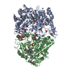
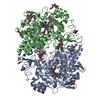
Deposited unit
A: Prostaglandin G/H synthase 2
B: Prostaglandin G/H synthase 2
hetero molecules


Theoretical massNumber of molelcules
Total (without water)131,00419
Polymers126,8232
Non-polymers4,18117
Water2,774154
1


  • Idetical with deposited unit
  • defined by author&software
TypeNameSymmetry operationNumber
identity operation1_555x,y,z1
Buried area11460 Å2
ΔGint-63 kcal/mol
Surface area41530 Å2
MethodPISA
Unit cell
Length a, b, c (Å)126.989, 149.422, 185.055
Angle α, β, γ (deg.)90.00, 90.00, 90.00
Int Tables number23
Space group name H-MI222

-
Components

-
Protein , 1 types, 2 molecules AB

#1: Protein Prostaglandin G/H synthase 2 / Cyclooxygenase-2 / COX-2 / PHS II / Prostaglandin H2 synthase 2 / PGHS-2 / Prostaglandin- ...Cyclooxygenase-2 / COX-2 / PHS II / Prostaglandin H2 synthase 2 / PGHS-2 / Prostaglandin-endoperoxide synthase 2


Mass: 63411.312 Da / Num. of mol.: 2
Source method: isolated from a genetically manipulated source
Source: (gene. exp.) Homo sapiens (human) / Gene: PTGS2, COX2 / Production host: Spodoptera frugiperda (fall armyworm)
References: UniProt: P35354, prostaglandin-endoperoxide synthase

-
Sugars , 2 types, 4 molecules

#2: Polysaccharide alpha-D-mannopyranose-(1-4)-2-acetamido-2-deoxy-beta-D-glucopyranose-(1-4)-2-acetamido-2-deoxy-beta- ...alpha-D-mannopyranose-(1-4)-2-acetamido-2-deoxy-beta-D-glucopyranose-(1-4)-2-acetamido-2-deoxy-beta-D-glucopyranose


Type: oligosaccharide / Mass: 586.542 Da / Num. of mol.: 2
Source method: isolated from a genetically manipulated source
DescriptorTypeProgram
DManpa1-4DGlcpNAcb1-4DGlcpNAcb1-Glycam Condensed SequenceGMML 1.0
WURCS=2.0/2,3,2/[a2122h-1b_1-5_2*NCC/3=O][a1122h-1a_1-5]/1-1-2/a4-b1_b4-c1WURCSPDB2Glycan 1.1.0
[]{[(4+1)][b-D-GlcpNAc]{[(4+1)][b-D-GlcpNAc]{[(4+1)][a-D-Manp]{}}}}LINUCSPDB-CARE
#5: Sugar ChemComp-NAG / 2-acetamido-2-deoxy-beta-D-glucopyranose / N-acetyl-beta-D-glucosamine / 2-acetamido-2-deoxy-beta-D-glucose / 2-acetamido-2-deoxy-D-glucose / 2-acetamido-2-deoxy-glucose / N-ACETYL-D-GLUCOSAMINE


Type: D-saccharide, beta linking / Mass: 221.208 Da / Num. of mol.: 2
Source method: isolated from a genetically manipulated source
Formula: C8H15NO6
IdentifierTypeProgram
DGlcpNAcbCONDENSED IUPAC CARBOHYDRATE SYMBOLGMML 1.0
N-acetyl-b-D-glucopyranosamineCOMMON NAMEGMML 1.0
b-D-GlcpNAcIUPAC CARBOHYDRATE SYMBOLPDB-CARE 1.0
GlcNAcSNFG CARBOHYDRATE SYMBOLGMML 1.0

-
Non-polymers , 6 types, 167 molecules

#3: Chemical ChemComp-RCX / Rofecoxib / Vioxx


Mass: 314.356 Da / Num. of mol.: 2
Source method: isolated from a genetically manipulated source
Formula: C17H14O4S / Comment: antiinflammatory*YM
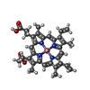
#4: Chemical ChemComp-COH / PROTOPORPHYRIN IX CONTAINING CO


Mass: 619.575 Da / Num. of mol.: 2
Source method: isolated from a genetically manipulated source
Formula: C34H32CoN4O4
#6: Chemical ChemComp-NH4 / AMMONIUM ION


Mass: 18.038 Da / Num. of mol.: 2 / Source method: obtained synthetically / Formula: H4N
#7: Chemical ChemComp-GOL / GLYCEROL / GLYCERIN / PROPANE-1,2,3-TRIOL


Mass: 92.094 Da / Num. of mol.: 1 / Source method: obtained synthetically / Formula: C3H8O3
#8: Chemical
ChemComp-PO4 / PHOSPHATE ION


Mass: 94.971 Da / Num. of mol.: 6 / Source method: obtained synthetically / Formula: PO4
#9: Water ChemComp-HOH / water


Mass: 18.015 Da / Num. of mol.: 154 / Source method: isolated from a natural source / Formula: H2O

-
Experimental details

-
Experiment

ExperimentMethod: X-RAY DIFFRACTION / Number of used crystals: 1

-
Sample preparation

CrystalDensity Matthews: 3.46 Å3/Da / Density % sol: 64.46 %
Crystal growTemperature: 296 K / Method: vapor diffusion, sitting drop / pH: 7 / Details: PEG400, ammonium phosphate, HEPES

-
Data collection

DiffractionMean temperature: 100 K
Diffraction sourceSource: SYNCHROTRON / Site: APS / Beamline: 23-ID-B / Wavelength: 1 Å
DetectorType: MARMOSAIC 300 mm CCD / Detector: CCD / Date: Nov 9, 2015
RadiationProtocol: SINGLE WAVELENGTH / Monochromatic (M) / Laue (L): M / Scattering type: x-ray
Radiation wavelengthWavelength: 1 Å / Relative weight: 1
ReflectionResolution: 2.69→30 Å / Num. obs: 48415 / % possible obs: 100 % / Redundancy: 7.1 % / Net I/σ(I): 17.9

-
Processing

Software
NameVersionClassification
PHENIX1.9_1692refinement
HKL-2000data reduction
HKL-2000data scaling
PHASERphasing
RefinementMethod to determine structure: MOLECULAR REPLACEMENT
Starting model: 3HS5
Resolution: 2.697→30.029 Å / SU ML: 0.29 / Cross valid method: FREE R-VALUE / σ(F): 1.35 / Phase error: 22.16 / Stereochemistry target values: ML
RfactorNum. reflection% reflection
Rfree0.2196 2219 4.89 %
Rwork0.1781 --
obs0.1801 45368 93.12 %
Solvent computationShrinkage radii: 0.9 Å / VDW probe radii: 1.11 Å / Solvent model: FLAT BULK SOLVENT MODEL
Refinement stepCycle: LAST / Resolution: 2.697→30.029 Å
ProteinNucleic acidLigandSolventTotal
Num. atoms8916 0 274 154 9344
Refine LS restraints
Refine-IDTypeDev idealNumber
X-RAY DIFFRACTIONf_bond_d0.0059485
X-RAY DIFFRACTIONf_angle_d1.27512901
X-RAY DIFFRACTIONf_dihedral_angle_d14.6563488
X-RAY DIFFRACTIONf_chiral_restr0.1331354
X-RAY DIFFRACTIONf_plane_restr0.0051658
LS refinement shell
Resolution (Å)Rfactor RfreeNum. reflection RfreeRfactor RworkNum. reflection RworkRefine-ID% reflection obs (%)
2.6968-2.75540.26371000.21031709X-RAY DIFFRACTION60
2.7554-2.81940.22861000.21981995X-RAY DIFFRACTION70
2.8194-2.88990.3055970.22812252X-RAY DIFFRACTION78
2.8899-2.9680.25761270.22262487X-RAY DIFFRACTION87
2.968-3.05520.26711510.21862717X-RAY DIFFRACTION95
3.0552-3.15370.26991430.22382826X-RAY DIFFRACTION99
3.1537-3.26630.3071520.21892888X-RAY DIFFRACTION100
3.2663-3.39690.23621510.20472872X-RAY DIFFRACTION100
3.3969-3.55130.22141410.1842910X-RAY DIFFRACTION100
3.5513-3.73820.20711400.17432892X-RAY DIFFRACTION100
3.7382-3.97190.20311610.1592867X-RAY DIFFRACTION100
3.9719-4.27780.20171530.15572879X-RAY DIFFRACTION100
4.2778-4.70680.19681450.14082924X-RAY DIFFRACTION100
4.7068-5.38450.18751590.13992913X-RAY DIFFRACTION100
5.3845-6.7710.22061580.18122959X-RAY DIFFRACTION100
6.771-30.03070.18641410.17613059X-RAY DIFFRACTION100
Refinement TLS params.

Method: refined / Refine-ID: X-RAY DIFFRACTION

IDL112)L122)L132)L222)L232)L332)S11 (Å °)S12 (Å °)S13 (Å °)S21 (Å °)S22 (Å °)S23 (Å °)S31 (Å °)S32 (Å °)S33 (Å °)T112)T122)T132)T222)T232)T332)Origin x (Å)Origin y (Å)Origin z (Å)
12.9137-0.6248-0.47012.1189-0.95580.64040.0596-0.11740.07390.69990.20790.9914-0.3098-0.5411-0.04530.44750.15860.17620.46280.10410.56547.101727.329239.8511
21.3350.6627-0.31032.1769-0.21242.2322-0.07430.3979-0.5106-0.3330.13470.24380.3078-0.1668-0.04370.2494-0.0691-0.07390.4282-0.17920.5512.35180.332325.6085
31.3584-0.0294-0.09751.8339-0.19050.8895-0.0516-0.0597-0.42090.25090.0266-0.52090.07630.12070.02160.22480.061-0.0540.1936-0.07050.440139.555310.24633.3473
41.1659-0.6201-0.83831.5643-0.20440.9875-0.2413-0.2617-0.67690.46520.1204-0.41730.11050.18840.00870.36150.015-0.1680.30410.05430.719839.41182.667939.7198
51.32680.0435-0.32292.2122-0.35241.4724-0.0201-0.1655-0.77550.4592-0.0108-0.08720.0678-0.0940.0830.27540.0181-0.06070.1917-0.07490.432527.79114.156338.8979
60.39560.35820.26381.36640.05321.0223-0.04850.5451-0.3179-0.7905-0.0477-0.13510.10670.0361-0.93770.8957-0.09750.10480.7615-0.36090.367536.171913.6108-3.135
72.2103-0.7780.68810.9102-0.71350.577-0.25650.34310.115-0.62960.1470.1080.0413-0.1546-0.11750.477-0.0457-0.20480.5253-0.07530.391522.439229.7766.0668
82.334-1.3292-0.61632.55931.69473.1842-0.20620.65640.1339-1.0301-0.0745-0.83770.02270.55340.29810.6813-0.12490.19550.7662-0.00990.471647.821434.5749-1.3182
91.45710.12-0.4172.4775-0.03071.4559-0.0940.16720.3858-0.04920.20590.0291-0.34690.0443-0.08430.3630.0231-0.12650.2291-0.00470.284629.917848.426121.48
100.8182-0.08170.25441.8595-0.25660.8701-0.09360.5510.2338-0.72580.08780.0777-0.19750.05350.02810.53-0.0715-0.07570.50690.02670.266830.525740.9285.1204
Refinement TLS group
IDRefine-IDRefine TLS-IDSelection details
1X-RAY DIFFRACTION1(chain A and resid 34:73)
2X-RAY DIFFRACTION2(chain A and resid 74:122)
3X-RAY DIFFRACTION3(chain A and resid 123:367)
4X-RAY DIFFRACTION4(chain A and resid 368:419)
5X-RAY DIFFRACTION5(chain A and resid 420:583)
6X-RAY DIFFRACTION6(chain B and resid 34:78)
7X-RAY DIFFRACTION7(chain B and resid 79:147)
8X-RAY DIFFRACTION8(chain B and resid 148:183)
9X-RAY DIFFRACTION9(chain B and resid 184:429)
10X-RAY DIFFRACTION10(chain B and resid 430:583)

+
About Yorodumi

-
News

-
Feb 9, 2022. New format data for meta-information of EMDB entries

New format data for meta-information of EMDB entries

  • Version 3 of the EMDB header file is now the official format.
  • The previous official version 1.9 will be removed from the archive.

Related info.:EMDB header

External links:wwPDB to switch to version 3 of the EMDB data model

-
Aug 12, 2020. Covid-19 info

Covid-19 info

URL: https://pdbj.org/emnavi/covid19.php

New page: Covid-19 featured information page in EM Navigator.

Related info.:Covid-19 info / Mar 5, 2020. Novel coronavirus structure data

+
Mar 5, 2020. Novel coronavirus structure data

Novel coronavirus structure data

Related info.:Yorodumi Speices / Aug 12, 2020. Covid-19 info

External links:COVID-19 featured content - PDBj / Molecule of the Month (242):Coronavirus Proteases

+
Jan 31, 2019. EMDB accession codes are about to change! (news from PDBe EMDB page)

EMDB accession codes are about to change! (news from PDBe EMDB page)

  • The allocation of 4 digits for EMDB accession codes will soon come to an end. Whilst these codes will remain in use, new EMDB accession codes will include an additional digit and will expand incrementally as the available range of codes is exhausted. The current 4-digit format prefixed with “EMD-” (i.e. EMD-XXXX) will advance to a 5-digit format (i.e. EMD-XXXXX), and so on. It is currently estimated that the 4-digit codes will be depleted around Spring 2019, at which point the 5-digit format will come into force.
  • The EM Navigator/Yorodumi systems omit the EMD- prefix.

Related info.:Q: What is EMD? / ID/Accession-code notation in Yorodumi/EM Navigator

External links:EMDB Accession Codes are Changing Soon! / Contact to PDBj

+
Jul 12, 2017. Major update of PDB

Major update of PDB

  • wwPDB released updated PDB data conforming to the new PDBx/mmCIF dictionary.
  • This is a major update changing the version number from 4 to 5, and with Remediation, in which all the entries are updated.
  • In this update, many items about electron microscopy experimental information are reorganized (e.g. em_software).
  • Now, EM Navigator and Yorodumi are based on the updated data.

External links:wwPDB Remediation / Enriched Model Files Conforming to OneDep Data Standards Now Available in the PDB FTP Archive

-
Yorodumi

Thousand views of thousand structures

  • Yorodumi is a browser for structure data from EMDB, PDB, SASBDB, etc.
  • This page is also the successor to EM Navigator detail page, and also detail information page/front-end page for Omokage search.
  • The word "yorodu" (or yorozu) is an old Japanese word meaning "ten thousand". "mi" (miru) is to see.

Related info.:EMDB / PDB / SASBDB / Comparison of 3 databanks / Yorodumi Search / Aug 31, 2016. New EM Navigator & Yorodumi / Yorodumi Papers / Jmol/JSmol / Function and homology information / Changes in new EM Navigator and Yorodumi

Read more