Mass: 18.015 Da / Num. of mol.: 14 / Source method: isolated from a natural source / Formula: H2O
-
Experimental details
-
Experiment
Experiment
Method: X-RAY DIFFRACTION / Number of used crystals: 1
-
Sample preparation
Crystal
Density Matthews: 2.57 Å3/Da / Density % sol: 52 %
Crystal grow
Temperature: 289 K / Method: vapor diffusion, sitting drop / pH: 7.4 Details: DAAO AT 2 MG/ML IN 50 MM SODIUM PHOSPHATE PH 6.6, 10 UM FAD INCUBATED WITH 5-FOLD EXCESS OF SEP-0374036 OVERNIGHT AT 277 K, CRYSTALLIZED AGAINST 13.6% PEG3350, 0.1 M TRIS PH 7.4, 150 MM ...Details: DAAO AT 2 MG/ML IN 50 MM SODIUM PHOSPHATE PH 6.6, 10 UM FAD INCUBATED WITH 5-FOLD EXCESS OF SEP-0374036 OVERNIGHT AT 277 K, CRYSTALLIZED AGAINST 13.6% PEG3350, 0.1 M TRIS PH 7.4, 150 MM POTASSIUM CITRATE TRIBASIC AND 20 MM GLYCEROL AS CRYO-PROTECTANT, CRYSTAL TRACKING ID 241709B5, UNIQUE PUCK ID XSS6-4, VAPOR DIFFUSION, SITTING DROP, temperature 289K
Resolution: 2.85→19.91 Å / Cor.coef. Fo:Fc: 0.912 / Cor.coef. Fo:Fc free: 0.873 / WRfactor Rfree: 0.2523 / WRfactor Rwork: 0.216 / FOM work R set: 0.7299 / SU B: 52.653 / SU ML: 0.433 / SU R Cruickshank DPI: 0.4135 / SU Rfree: 0.436 / Cross valid method: THROUGHOUT / σ(F): 0 / ESU R Free: 0.436 / Stereochemistry target values: MAXIMUM LIKELIHOOD Details: U VALUES : WITH TLS ADDED HYDROGENS HAVE BEEN ADDED IN THE RIDING POSITIONS
Rfactor
Num. reflection
% reflection
Selection details
Rfree
0.287
1000
5.1 %
RANDOM
Rwork
0.248
-
-
-
obs
0.25
19539
99.13 %
-
Solvent computation
Ion probe radii: 0.8 Å / Shrinkage radii: 0.8 Å / VDW probe radii: 1.2 Å / Solvent model: MASK
In the structure databanks used in Yorodumi, some data are registered as the other names, "COVID-19 virus" and "2019-nCoV". Here are the details of the virus and the list of structure data.
Jan 31, 2019. EMDB accession codes are about to change! (news from PDBe EMDB page)
EMDB accession codes are about to change! (news from PDBe EMDB page)
The allocation of 4 digits for EMDB accession codes will soon come to an end. Whilst these codes will remain in use, new EMDB accession codes will include an additional digit and will expand incrementally as the available range of codes is exhausted. The current 4-digit format prefixed with “EMD-” (i.e. EMD-XXXX) will advance to a 5-digit format (i.e. EMD-XXXXX), and so on. It is currently estimated that the 4-digit codes will be depleted around Spring 2019, at which point the 5-digit format will come into force.
The EM Navigator/Yorodumi systems omit the EMD- prefix.
Related info.:Q: What is EMD? / ID/Accession-code notation in Yorodumi/EM Navigator
Yorodumi is a browser for structure data from EMDB, PDB, SASBDB, etc.
This page is also the successor to EM Navigator detail page, and also detail information page/front-end page for Omokage search.
The word "yorodu" (or yorozu) is an old Japanese word meaning "ten thousand". "mi" (miru) is to see.
Related info.:EMDB / PDB / SASBDB / Comparison of 3 databanks / Yorodumi Search / Aug 31, 2016. New EM Navigator & Yorodumi / Yorodumi Papers / Jmol/JSmol / Function and homology information / Changes in new EM Navigator and Yorodumi
 Movie
Movie Controller

 Yorodumi
 Open data
 Basic information
 Components
 Keywords
 Function and homology information
 Homo sapiens (human)
 X-RAY DIFFRACTION /
  Authors
 Citation
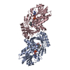
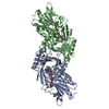
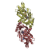
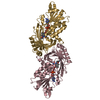
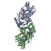
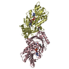
 Structure visualization
 Downloads & links
 Other downloads


 PDBj
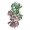
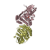
PDBj Assembly
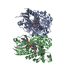


 Mass: 22.990 Da / Num. of mol.: 2 / Source method: obtained synthetically / Formula: Na
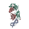
 Mass: 785.550 Da / Num. of mol.: 2 / Source method: obtained synthetically / Formula: C27H33N9O15P2 / Comment: FAD*YM
 Mass: 310.301 Da / Num. of mol.: 2 / Source method: obtained synthetically / Formula: C18H14O5
 Mass: 18.015 Da / Num. of mol.: 14 / Source method: isolated from a natural source / Formula: H2O
 Sample preparation
 / Beamline: BL7-1 / Wavelength: 1.12709 / Wavelength: 1.12709 Å
 Processing