 Movie
Movie Controller

[English] 日本語
 Yorodumi
- PDB-4kt0: Crystal structure of a virus like photosystem I from the cyanobac... -
+ Open data

- Basic information
| Entry | Database: PDB / ID: 4kt0 | |||||||||
|---|---|---|---|---|---|---|---|---|---|---|
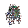
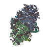
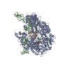
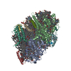
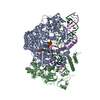
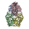
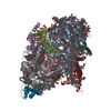
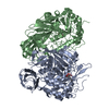
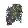
| Title | Crystal structure of a virus like photosystem I from the cyanobacterium Synechocystis PCC 6803 | |||||||||
 Components Components |
| |||||||||
 Keywords Keywords | ELECTRON TRANSPORT / photosynthetic reaction center / membrane complex / plastocyanin / cytochrome C6 / Ferredoxin | |||||||||
| Function / homology |  Function and homology information Function and homology informationthylakoid membrane / photosystem I reaction center / photosystem I / photosynthetic electron transport in photosystem I / photosystem I / plasma membrane-derived thylakoid membrane / chlorophyll binding / membrane => GO:0016020 / photosynthesis / 4 iron, 4 sulfur cluster binding ...thylakoid membrane / photosystem I reaction center / photosystem I / photosynthetic electron transport in photosystem I / photosystem I / plasma membrane-derived thylakoid membrane / chlorophyll binding / membrane => GO:0016020 / photosynthesis / 4 iron, 4 sulfur cluster binding / electron transfer activity / oxidoreductase activity / magnesium ion binding / metal ion binding / plasma membrane Similarity search - Function | |||||||||
| Biological species |  | |||||||||
| Method |  X-RAY DIFFRACTION / SYNCHROTRON / MOLECULAR REPLACEMENT / Resolution: 2.8 Å X-RAY DIFFRACTION / SYNCHROTRON / MOLECULAR REPLACEMENT / Resolution: 2.8 Å | |||||||||
 Authors Authors | Mazor, Y. / Nataf, D. / Toporik, H. / Nelson, N. | |||||||||
 Citation Citation | Journal: Elife / Year: 2014 Title: Crystal structures of virus-like photosystem I complexes from the mesophilic cyanobacterium Synechocystis PCC 6803. Authors: Mazor, Y. / Nataf, D. / Toporik, H. / Nelson, N. | |||||||||
| History |
|
- Structure visualization
| Structure viewer | Molecule: MolmilJmol/JSmol |
|---|
- Downloads & links
-Download
| PDBx/mmCIF format | 4kt0.cif.gz | 1.1 MB | Display | PDBx/mmCIF format |
|---|---|---|---|---|
| PDB format | pdb4kt0.ent.gz | 972.9 KB | Display | PDB format |
| PDBx/mmJSON format | 4kt0.json.gz | Tree view | PDBx/mmJSON format | |
| Others |  Other downloads Other downloads |
-Validation report
| Arichive directory | https://data.pdbj.org/pub/pdb/validation_reports/kt/4kt0ftp://data.pdbj.org/pub/pdb/validation_reports/kt/4kt0 | HTTPS FTP |
|---|
-Related structure data
| Related structure data |  4l6vC 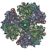 1jb0S S: Starting model for refinement C: citing same article ( |
|---|---|
| Similar structure data |
-Links
 PDBj
PDBj


- Assembly
| Deposited unit | 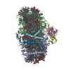
| ||||||||
|---|---|---|---|---|---|---|---|---|---|
| 1 |
| ||||||||
| Unit cell |
| ||||||||
| Details | AUTHOR STATED THAT THERE IS NO QUATERNARY STRUCTURE FOR THIS ENTRY. PISA FAILED TO GENERATE ASSEMBLY. THEREFORE THE ASU IS INDICATED AS ASSEMBLY TEMPORARILY. |
-Components
-Photosystem I P700 chlorophyll a apoprotein ... , 2 types, 2 molecules AB
| #1: Protein | Mass: 83036.398 Da / Num. of mol.: 1 Source method: isolated from a genetically manipulated source Details: 10HIS tag was added to the C terminus of PsaL. The mutated chain is lost during purification. Source: (gene. exp.) References: UniProt: L8AHT3, UniProt: P29254*PLUS, photosystem I |
|---|---|
| #2: Protein | Mass: 81369.531 Da / Num. of mol.: 1 Source method: isolated from a genetically manipulated source Source: (gene. exp.) References: UniProt: L8AIC0, UniProt: P29255*PLUS, photosystem I |
-Photosystem I subunit ... , 2 types, 2 molecules DF
| #4: Protein | Mass: 15663.749 Da / Num. of mol.: 1 Source method: isolated from a genetically manipulated source Source: (gene. exp.) References: UniProt: L8AFM8, UniProt: P19569*PLUS, photosystem I |
|---|---|
| #6: Protein | Mass: 18267.082 Da / Num. of mol.: 1 Source method: isolated from a genetically manipulated source Source: (gene. exp.) References: UniProt: L8AII8, UniProt: P29256*PLUS, photosystem I |
-Photosystem I reaction center subunit ... , 4 types, 4 molecules EJKM
| #5: Protein | Mass: 8154.086 Da / Num. of mol.: 1 Source method: isolated from a genetically manipulated source Source: (gene. exp.) References: UniProt: L8ASH8, UniProt: P12975*PLUS, photosystem I |
|---|---|
| #7: Protein/peptide | Mass: 4535.415 Da / Num. of mol.: 1 Source method: isolated from a genetically manipulated source Source: (gene. exp.) References: UniProt: L8AGL9, UniProt: Q55329*PLUS, photosystem I |
| #8: Protein | Mass: 13732.274 Da / Num. of mol.: 1 Source method: isolated from a genetically manipulated source Source: (gene. exp.) References: UniProt: L8APJ0, UniProt: P74564*PLUS, photosystem I |
| #9: Protein/peptide | Mass: 3382.063 Da / Num. of mol.: 1 Source method: isolated from a genetically manipulated source Source: (gene. exp.) References: UniProt: L8ADF9, UniProt: P72986*PLUS, photosystem I |
-Protein / Sugars , 2 types, 3 molecules C
| #16: Sugar |  Type: D-saccharide / Mass: 510.615 Da / Num. of mol.: 2 / Source method: obtained synthetically / Formula: C24H46O11 / Comment: detergent*YM Type: D-saccharide / Mass: 510.615 Da / Num. of mol.: 2 / Source method: obtained synthetically / Formula: C24H46O11 / Comment: detergent*YM#3: Protein | | Mass: 8837.261 Da / Num. of mol.: 1 Source method: isolated from a genetically manipulated source Source: (gene. exp.) References: UniProt: L8AST2, UniProt: P32422*PLUS, photosystem I |
|---|
-Non-polymers , 9 types, 149 molecules 


| #10: Chemical |  Mass: 450.696 Da / Num. of mol.: 2 / Source method: obtained synthetically / Formula: C31H46O2 Mass: 450.696 Da / Num. of mol.: 2 / Source method: obtained synthetically / Formula: C31H46O2#11: Chemical |  Mass: 351.640 Da / Num. of mol.: 3 / Source method: obtained synthetically / Formula: Fe4S4 Mass: 351.640 Da / Num. of mol.: 3 / Source method: obtained synthetically / Formula: Fe4S4#12: Chemical | ChemComp-LHG /  Mass: 722.970 Da / Num. of mol.: 4 / Source method: obtained synthetically / Formula: C38H75O10P / Comment: phospholipid*YM Mass: 722.970 Da / Num. of mol.: 4 / Source method: obtained synthetically / Formula: C38H75O10P / Comment: phospholipid*YM#13: Chemical |  Mass: 893.489 Da / Num. of mol.: 2 / Source method: obtained synthetically / Formula: C55H72MgN4O5 Mass: 893.489 Da / Num. of mol.: 2 / Source method: obtained synthetically / Formula: C55H72MgN4O5#14: Chemical | ChemComp-BCR /  Mass: 536.873 Da / Num. of mol.: 17 / Source method: obtained synthetically / Formula: C40H56 Mass: 536.873 Da / Num. of mol.: 17 / Source method: obtained synthetically / Formula: C40H56#15: Chemical | ChemComp-CLA /  Mass: 893.489 Da / Num. of mol.: 90 / Source method: obtained synthetically / Formula: C55H72MgN4O5 Mass: 893.489 Da / Num. of mol.: 90 / Source method: obtained synthetically / Formula: C55H72MgN4O5#17: Chemical | ChemComp-LMG / |  Mass: 787.158 Da / Num. of mol.: 1 / Source method: obtained synthetically / Formula: C45H86O10 Mass: 787.158 Da / Num. of mol.: 1 / Source method: obtained synthetically / Formula: C45H86O10#18: Chemical | ChemComp-CL / |  Mass: 35.453 Da / Num. of mol.: 1 / Source method: obtained synthetically / Formula: Cl Mass: 35.453 Da / Num. of mol.: 1 / Source method: obtained synthetically / Formula: Cl#19: Water | ChemComp-HOH / | Mass: 18.015 Da / Num. of mol.: 29 / Source method: isolated from a natural source / Formula: H2O |
|---|
-Details
| Has protein modification | Y |
|---|
-Experimental details
-Experiment
| Experiment | Method: X-RAY DIFFRACTION / Number of used crystals: 1 |
|---|
- Sample preparation
| Crystal | Density Matthews: 3.94 Å3/Da / Density % sol: 68.75 % |
|---|---|
| Crystal grow | Temperature: 278 K / Method: vapor diffusion, sitting drop / pH: 8.5 Details: 4.5% PEG3350, 30mM Tricine-NaOH pH8, 55mM NaCl, 50mM Glycine, 0.005% Nonyl- beta -D-Maltoside., pH 8.5, VAPOR DIFFUSION, SITTING DROP, temperature 278K |
-Data collection
| Diffraction | Mean temperature: 100 K |
|---|---|
| Diffraction source | Source: SYNCHROTRON / Site: ESRF  / Beamline: ID29 / Wavelength: 0.97625 Å / Beamline: ID29 / Wavelength: 0.97625 Å |
| Detector | Type: DECTRIS PILATUS 6M / Detector: PIXEL / Date: Feb 16, 2013 |
| Radiation | Protocol: SINGLE WAVELENGTH / Monochromatic (M) / Laue (L): M / Scattering type: x-ray |
| Radiation wavelength | Wavelength: 0.97625 Å / Relative weight: 1 |
| Reflection | Resolution: 2.8→30 Å / Num. all: 92450 / Num. obs: 91895 / % possible obs: 99.4 % / Observed criterion σ(F): 1 / Observed criterion σ(I): 1 / Redundancy: 4.6 % / Biso Wilson estimate: 61.4 Å2 / Rmerge(I) obs: 0.139 / Net I/σ(I): 7.9 |
| Reflection shell | Resolution: 2.8→2.95 Å / Redundancy: 4.1 % / Rmerge(I) obs: 2.382 / Mean I/σ(I) obs: 1.1 |
- Processing
| Software |
| |||||||||||||||||||||||||||||||||||||||||||||||||||||||||||||||||||||||||||||||||||||||||||||||||||||||||||||||||||||||||||||||||||||||||||||||||||||||||||||||||||||||||||||||||||||||||||||||||||||||||||||||||||||||||
|---|---|---|---|---|---|---|---|---|---|---|---|---|---|---|---|---|---|---|---|---|---|---|---|---|---|---|---|---|---|---|---|---|---|---|---|---|---|---|---|---|---|---|---|---|---|---|---|---|---|---|---|---|---|---|---|---|---|---|---|---|---|---|---|---|---|---|---|---|---|---|---|---|---|---|---|---|---|---|---|---|---|---|---|---|---|---|---|---|---|---|---|---|---|---|---|---|---|---|---|---|---|---|---|---|---|---|---|---|---|---|---|---|---|---|---|---|---|---|---|---|---|---|---|---|---|---|---|---|---|---|---|---|---|---|---|---|---|---|---|---|---|---|---|---|---|---|---|---|---|---|---|---|---|---|---|---|---|---|---|---|---|---|---|---|---|---|---|---|---|---|---|---|---|---|---|---|---|---|---|---|---|---|---|---|---|---|---|---|---|---|---|---|---|---|---|---|---|---|---|---|---|---|---|---|---|---|---|---|---|---|---|---|---|---|---|---|---|---|
| Refinement | Method to determine structure: MOLECULAR REPLACEMENT Starting model: PDB entry 1JB0 with truncated side chains and chlorophyll tails Resolution: 2.8→29.977 Å / SU ML: 0.4 / Isotropic thermal model: single parameter per residue. / σ(F): 1.35 / Phase error: 28.46 / Stereochemistry target values: ML
| |||||||||||||||||||||||||||||||||||||||||||||||||||||||||||||||||||||||||||||||||||||||||||||||||||||||||||||||||||||||||||||||||||||||||||||||||||||||||||||||||||||||||||||||||||||||||||||||||||||||||||||||||||||||||
| Solvent computation | Shrinkage radii: 0.9 Å / VDW probe radii: 1.11 Å / Solvent model: FLAT BULK SOLVENT MODEL | |||||||||||||||||||||||||||||||||||||||||||||||||||||||||||||||||||||||||||||||||||||||||||||||||||||||||||||||||||||||||||||||||||||||||||||||||||||||||||||||||||||||||||||||||||||||||||||||||||||||||||||||||||||||||
| Displacement parameters | Biso mean: 94.61 Å2 | |||||||||||||||||||||||||||||||||||||||||||||||||||||||||||||||||||||||||||||||||||||||||||||||||||||||||||||||||||||||||||||||||||||||||||||||||||||||||||||||||||||||||||||||||||||||||||||||||||||||||||||||||||||||||
| Refinement step | Cycle: LAST / Resolution: 2.8→29.977 Å
| |||||||||||||||||||||||||||||||||||||||||||||||||||||||||||||||||||||||||||||||||||||||||||||||||||||||||||||||||||||||||||||||||||||||||||||||||||||||||||||||||||||||||||||||||||||||||||||||||||||||||||||||||||||||||
| Refine LS restraints |
| |||||||||||||||||||||||||||||||||||||||||||||||||||||||||||||||||||||||||||||||||||||||||||||||||||||||||||||||||||||||||||||||||||||||||||||||||||||||||||||||||||||||||||||||||||||||||||||||||||||||||||||||||||||||||
| LS refinement shell |
| |||||||||||||||||||||||||||||||||||||||||||||||||||||||||||||||||||||||||||||||||||||||||||||||||||||||||||||||||||||||||||||||||||||||||||||||||||||||||||||||||||||||||||||||||||||||||||||||||||||||||||||||||||||||||
| Refinement TLS params. | Method: refined / Origin x: -9.005 Å / Origin y: -7.8261 Å / Origin z: 21.7491 Å
| |||||||||||||||||||||||||||||||||||||||||||||||||||||||||||||||||||||||||||||||||||||||||||||||||||||||||||||||||||||||||||||||||||||||||||||||||||||||||||||||||||||||||||||||||||||||||||||||||||||||||||||||||||||||||
| Refinement TLS group | Selection details: chain A or chain B or chain C or chain D or chain E or chain F or chain J or chain K or chain M |
