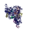
 Movie
Movie Controller


+ Open data

- Basic information
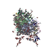
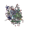
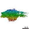
| Entry | Database: PDB / ID: 3lw5 | |||||||||
|---|---|---|---|---|---|---|---|---|---|---|
| Title | Improved model of plant photosystem I | |||||||||
 Components Components |
| |||||||||
 Keywords Keywords | PHOTOSYNTHESIS / Electron transfer / Membrane proteins / Large / 2 Complexes / Chlorophyll / Chloroplast / Chromophore / Electron transport / Iron / Iron-sulfur / Magnesium / Membrane / Metal-binding / Photosystem I / Thylakoid / Transmembrane / Transport | |||||||||
| Function / homology |  Function and homology information Function and homology informationchloroplast photosystem I / photosynthesis, light harvesting in photosystem I / photosynthesis, light harvesting / chloroplast thylakoid lumen / photosystem I reaction center / photosystem I / photosynthetic electron transport in photosystem I / photosystem I / photosystem II / chlorophyll binding ...chloroplast photosystem I / photosynthesis, light harvesting in photosystem I / photosynthesis, light harvesting / chloroplast thylakoid lumen / photosystem I reaction center / photosystem I / photosynthetic electron transport in photosystem I / photosystem I / photosystem II / chlorophyll binding / chloroplast thylakoid membrane / photosynthesis / phosphoprotein binding / 4 iron, 4 sulfur cluster binding / electron transfer activity / oxidoreductase activity / protein domain specific binding / magnesium ion binding / metal ion binding Similarity search - Function | |||||||||
| Biological species |  Pisum sativum (garden pea) Pisum sativum (garden pea)unidentified (others) | |||||||||
| Method |  X-RAY DIFFRACTION / SYNCHROTRON / MOLECULAR REPLACEMENT / molecular replacement / Resolution: 3.3 Å X-RAY DIFFRACTION / SYNCHROTRON / MOLECULAR REPLACEMENT / molecular replacement / Resolution: 3.3 Å | |||||||||
 Authors Authors | Nelson, N. / Toporik, H. | |||||||||
 Citation Citation | Journal: J.Biol.Chem. / Year: 2010 Title: Structure determination and improved model of plant photosystem I Authors: Amunts, A. / Toporik, H. / Borovikova, A. / Nelson, N. | |||||||||
| History |
|
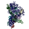
- Structure visualization
| Structure viewer | Molecule: MolmilJmol/JSmol |
|---|
- Downloads & links
-Download
| PDBx/mmCIF format | 3lw5.cif.gz | 939 KB | Display | PDBx/mmCIF format |
|---|---|---|---|---|
| PDB format | pdb3lw5.ent.gz | 796.2 KB | Display | PDB format |
| PDBx/mmJSON format | 3lw5.json.gz | Tree view | PDBx/mmJSON format | |
| Others |  Other downloads Other downloads |
-Validation report
| Arichive directory | https://data.pdbj.org/pub/pdb/validation_reports/lw/3lw5ftp://data.pdbj.org/pub/pdb/validation_reports/lw/3lw5 | HTTPS FTP |
|---|
-Related structure data
| Related structure data |  2wscSC 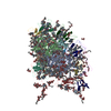 2wseC  2wsfC S: Starting model for refinement C: citing same article ( |
|---|---|
| Similar structure data |

-Links
 PDBj
PDBj


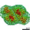
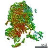
- Assembly
| Deposited unit | 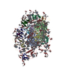
| ||||||||
|---|---|---|---|---|---|---|---|---|---|
| 1 |
| ||||||||
| Unit cell |
|
-Components
-Photosystem I P700 chlorophyll a apoprotein ... , 2 types, 2 molecules AB
| #1: Protein | Mass: 82022.758 Da / Num. of mol.: 1 / Fragment: residues 21-758 / Source method: isolated from a natural source / Source: (natural) Pisum sativum (garden pea) / References: UniProt: P05310 |
|---|---|
| #2: Protein | Mass: 82359.766 Da / Num. of mol.: 1 / Source method: isolated from a natural source / Source: (natural) Pisum sativum (garden pea) / References: UniProt: P05311 |
-Protein , 5 types, 5 molecules CNR12
| #3: Protein | Mass: 8991.474 Da / Num. of mol.: 1 / Source method: isolated from a natural source / Source: (natural) Pisum sativum (garden pea) / References: UniProt: P10793 |
|---|---|
| #13: Protein | Mass: 9767.049 Da / Num. of mol.: 1 / Source method: isolated from a natural source / Source: (natural) Pisum sativum (garden pea) / References: UniProt: E1C9K7*PLUS |
| #14: Protein | Mass: 4528.573 Da / Num. of mol.: 1 / Source method: isolated from a natural source / Source: (natural) unidentified (others) |
| #15: Protein | Mass: 18616.328 Da / Num. of mol.: 1 / Source method: isolated from a natural source / Source: (natural) Pisum sativum (garden pea) / References: UniProt: E1C9L2*PLUS |
| #16: Protein | Mass: 19528.191 Da / Num. of mol.: 1 / Fragment: residues 94-269 / Source method: isolated from a natural source / Source: (natural) Pisum sativum (garden pea) / References: UniProt: Q41038 |
-Putative uncharacterized ... , 5 types, 5 molecules DEGHL
| #4: Protein | Mass: 15557.807 Da / Num. of mol.: 1 / Source method: isolated from a natural source / Source: (natural) Pisum sativum (garden pea) / References: UniProt: E1C9K8*PLUS |
|---|---|
| #5: Protein | Mass: 7270.221 Da / Num. of mol.: 1 / Source method: isolated from a natural source / Source: (natural) Pisum sativum (garden pea) / References: UniProt: E1C9K6*PLUS |
| #7: Protein | Mass: 10456.749 Da / Num. of mol.: 1 / Source method: isolated from a natural source / Source: (natural) Pisum sativum (garden pea) / References: UniProt: P20120*PLUS |
| #8: Protein | Mass: 7314.132 Da / Num. of mol.: 1 / Source method: isolated from a natural source / Source: (natural) Pisum sativum (garden pea) / References: UniProt: E1C9K9*PLUS |
| #12: Protein | Mass: 17059.578 Da / Num. of mol.: 1 / Source method: isolated from a natural source / Source: (natural) Pisum sativum (garden pea) / References: UniProt: E1C9L1*PLUS |
-Photosystem I reaction center subunit ... , 4 types, 4 molecules FIJK
| #6: Protein | Mass: 17299.033 Da / Num. of mol.: 1 / Source method: isolated from a natural source / Source: (natural) Pisum sativum (garden pea) / References: UniProt: P12355*PLUS |
|---|---|
| #9: Protein/peptide | Mass: 3270.042 Da / Num. of mol.: 1 / Fragment: residues 1-30 / Source method: isolated from a natural source / Source: (natural) Pisum sativum (garden pea) / References: UniProt: P17227 |
| #10: Protein/peptide | Mass: 4793.689 Da / Num. of mol.: 1 / Source method: isolated from a natural source / Source: (natural) Pisum sativum (garden pea) / References: UniProt: E1C9L0*PLUS |
| #11: Protein | Mass: 8462.847 Da / Num. of mol.: 1 / Source method: isolated from a natural source / Source: (natural) Pisum sativum (garden pea) / References: UniProt: E1C9L3*PLUS |
-Chlorophyll a-b binding protein ... , 2 types, 2 molecules 34
| #17: Protein | Mass: 18723.426 Da / Num. of mol.: 1 / Fragment: residues 84-255 / Source method: isolated from a natural source / Source: (natural) Pisum sativum (garden pea) / References: UniProt: Q32904 |
|---|---|
| #18: Protein | Mass: 18704.330 Da / Num. of mol.: 1 / Fragment: residues 81-246 / Source method: isolated from a natural source / Source: (natural) Pisum sativum (garden pea) / References: UniProt: Q9SQL2 |
-Sugars , 1 types, 53 molecules 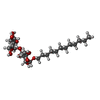
| #22: Sugar | ChemComp-LMU /  Type: D-saccharide / Mass: 510.615 Da / Num. of mol.: 53 / Source method: obtained synthetically / Formula: C24H46O11 / Comment: detergent*YM Type: D-saccharide / Mass: 510.615 Da / Num. of mol.: 53 / Source method: obtained synthetically / Formula: C24H46O11 / Comment: detergent*YM |
|---|
-Non-polymers , 5 types, 199 molecules 


| #19: Chemical | ChemComp-CLA /  Mass: 893.489 Da / Num. of mol.: 174 / Source method: obtained synthetically / Formula: C55H72MgN4O5 Mass: 893.489 Da / Num. of mol.: 174 / Source method: obtained synthetically / Formula: C55H72MgN4O5#20: Chemical |  Mass: 450.696 Da / Num. of mol.: 2 / Source method: obtained synthetically / Formula: C31H46O2 Mass: 450.696 Da / Num. of mol.: 2 / Source method: obtained synthetically / Formula: C31H46O2#21: Chemical | ChemComp-BCR /  Mass: 536.873 Da / Num. of mol.: 19 / Source method: obtained synthetically / Formula: C40H56 Mass: 536.873 Da / Num. of mol.: 19 / Source method: obtained synthetically / Formula: C40H56#23: Chemical |  Mass: 351.640 Da / Num. of mol.: 3 / Source method: obtained synthetically / Formula: Fe4S4 Mass: 351.640 Da / Num. of mol.: 3 / Source method: obtained synthetically / Formula: Fe4S4#24: Chemical | ChemComp-LMG / |  Mass: 787.158 Da / Num. of mol.: 1 / Source method: obtained synthetically / Formula: C45H86O10 Mass: 787.158 Da / Num. of mol.: 1 / Source method: obtained synthetically / Formula: C45H86O10 |
|---|
-Details
| Has protein modification | Y |
|---|---|
| Sequence details | MISSING ALANINE OF THIS POSITION IN CHAIN 4 IS CORRECT BECAUSE IN ALL THE OTHER PLANTS INCLUDING ...MISSING ALANINE OF THIS POSITION IN CHAIN 4 IS CORRECT BECAUSE IN ALL THE OTHER PLANTS INCLUDING ARABIDOPSI |
-Experimental details
-Experiment
| Experiment | Method: X-RAY DIFFRACTION / Number of used crystals: 1 |
|---|
- Sample preparation
| Crystal | Density Matthews: 4.16 Å3/Da / Density % sol: 70.43 % |
|---|---|
| Crystal grow | Temperature: 277 K / Method: vapor diffusion, sitting drop / pH: 6 Details: 22.5mM MES BIS-TRIS, 10mM SUCCINIC ACID, 6% PEG6000, 0.015% DTM, VAPOR DIFFUSION, SITTING DROP, temperature 277K |
-Data collection
| Diffraction | Mean temperature: 100 K |
|---|---|
| Diffraction source | Source: SYNCHROTRON / Site: ESRF  / Beamline: ID23-2 / Wavelength: 0.873 Å / Beamline: ID23-2 / Wavelength: 0.873 Å |
| Detector | Type: MARRESEARCH / Detector: CCD / Date: Jan 1, 2009 / Details: MIRRORS |
| Radiation | Monochromator: SI(111) / Protocol: SINGLE WAVELENGTH / Monochromatic (M) / Laue (L): M / Scattering type: x-ray |
| Radiation wavelength | Wavelength: 0.873 Å / Relative weight: 1 |
| Reflection | Resolution: 3.3→30 Å / Num. obs: 82447 |
-Phasing
| Phasing | Method: molecular replacement | |||||||||
|---|---|---|---|---|---|---|---|---|---|---|
| Phasing MR |
|
- Processing
| Software |
| |||||||||||||||||||||||||||||||||||||||||||||||||||||||||||||||||
|---|---|---|---|---|---|---|---|---|---|---|---|---|---|---|---|---|---|---|---|---|---|---|---|---|---|---|---|---|---|---|---|---|---|---|---|---|---|---|---|---|---|---|---|---|---|---|---|---|---|---|---|---|---|---|---|---|---|---|---|---|---|---|---|---|---|---|
| Refinement | Method to determine structure: MOLECULAR REPLACEMENT Starting model: 2WSC Resolution: 3.3→30 Å / Cor.coef. Fo:Fc: 0.792 / Cor.coef. Fo:Fc free: 0.743 / Occupancy max: 1 / Occupancy min: 1 / SU B: 50.537 / SU ML: 0.884 / Cross valid method: THROUGHOUT / σ(F): 0 / ESU R Free: 0.857 / Stereochemistry target values: MAXIMUM LIKELIHOOD Details: HYDROGENS HAVE BEEN ADDED IN THE RIDING POSITIONS U VALUES: REFINED INDIVIDUALLY
| |||||||||||||||||||||||||||||||||||||||||||||||||||||||||||||||||
| Displacement parameters | Biso max: 75.43 Å2 / Biso mean: 23.073 Å2 / Biso min: 2 Å2
| |||||||||||||||||||||||||||||||||||||||||||||||||||||||||||||||||
| Refinement step | Cycle: LAST / Resolution: 3.3→30 Å
| |||||||||||||||||||||||||||||||||||||||||||||||||||||||||||||||||
| Refine LS restraints |
| |||||||||||||||||||||||||||||||||||||||||||||||||||||||||||||||||
| LS refinement shell | Resolution: 3.3→3.385 Å / Total num. of bins used: 20
|
