 Movie
Movie Controller

[English] 日本語
 Yorodumi
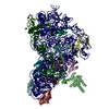
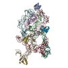
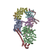
- EMDB-30511: Structural insights into a dimeric Psb27-photosystem II complex f... -
+ Open data

- Basic information
| Entry | Database: EMDB / ID: EMD-30511 | |||||||||
|---|---|---|---|---|---|---|---|---|---|---|
| Title | Structural insights into a dimeric Psb27-photosystem II complex from a cyanobacterium Thermosynechococcus vulcanus | |||||||||
 Map data Map data | PSII-Psb27 | |||||||||
 Sample Sample |
| |||||||||
 Keywords Keywords | dimeric photosystem II (PSII)-Psb27 complex from a cyanobacterium Thermosynechococcus vulcanus / PLANT PROTEIN | |||||||||
| Function / homology |  Function and homology information Function and homology informationoxygen evolving activity / photosystem II stabilization / photosystem II reaction center / photosystem II / photosynthetic electron transport chain / oxidoreductase activity, acting on diphenols and related substances as donors, oxygen as acceptor / response to herbicide / photosystem II / plasma membrane-derived thylakoid membrane / photosynthetic electron transport in photosystem II ...oxygen evolving activity / photosystem II stabilization / photosystem II reaction center / photosystem II / photosynthetic electron transport chain / oxidoreductase activity, acting on diphenols and related substances as donors, oxygen as acceptor / response to herbicide / photosystem II / plasma membrane-derived thylakoid membrane / photosynthetic electron transport in photosystem II / chlorophyll binding / photosynthesis, light reaction / phosphate ion binding / photosynthesis / electron transfer activity / protein stabilization / iron ion binding / heme binding / metal ion binding Similarity search - Function | |||||||||
| Biological species |  Thermosynechococcus vulcanus (bacteria) Thermosynechococcus vulcanus (bacteria) | |||||||||
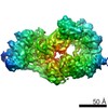
| Method | single particle reconstruction / cryo EM / Resolution: 3.78 Å | |||||||||
 Authors Authors | Pi X / Huang G | |||||||||
 Citation Citation | Journal: Proc Natl Acad Sci U S A / Year: 2021 Title: Structural insights into a dimeric Psb27-photosystem II complex from a cyanobacterium . Authors: Guoqiang Huang / Yanan Xiao / Xiong Pi / Liang Zhao / Qingjun Zhu / Wenda Wang / Tingyun Kuang / Guangye Han / Sen-Fang Sui / Jian-Ren Shen /   Abstract: Photosystem II (PSII) is a multisubunit pigment-protein complex and catalyzes light-driven water oxidation, leading to the conversion of light energy into chemical energy and the release of molecular ...Photosystem II (PSII) is a multisubunit pigment-protein complex and catalyzes light-driven water oxidation, leading to the conversion of light energy into chemical energy and the release of molecular oxygen. Psb27 is a small thylakoid lumen-localized protein known to serve as an assembly factor for the biogenesis and repair of the PSII complex. The exact location and binding fashion of Psb27 in the intermediate PSII remain elusive. Here, we report the structure of a dimeric Psb27-PSII complex purified from a deletion mutant (ΔPsbV) of the cyanobacterium , solved by cryo-electron microscopy. Our structure showed that Psb27 is associated with CP43 at the luminal side, with specific interactions formed between Helix 2 and Helix 3 of Psb27 and a loop region between Helix 3 and Helix 4 of CP43 (loop C) as well as the large, lumen-exposed and hydrophilic E-loop of CP43. The binding of Psb27 imposes some conflicts with the N-terminal region of PsbO and also induces some conformational changes in CP43, CP47, and D2. This makes PsbO unable to bind in the Psb27-PSII. Conformational changes also occurred in D1, PsbE, PsbF, and PsbZ; this, together with the conformational changes occurred in CP43, CP47, and D2, may prevent the binding of PsbU and induce dissociation of PsbJ. This structural information provides important insights into the regulation mechanism of Psb27 in the biogenesis and repair of PSII. | |||||||||
| History |
|
- Structure visualization
| Movie |
Movie viewer |
|---|---|
| Structure viewer | EM map: SurfViewMolmilJmol/JSmol |
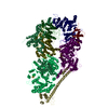
| Supplemental images |

- Downloads & links
-EMDB archive
| Map data | emd_30511.map.gz | 48.3 MB | EMDB map data format | |
|---|---|---|---|---|
| Header (meta data) | emd-30511-v30.xmlemd-30511.xml | 33.7 KB 33.7 KB | Display Display | EMDB header |
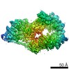
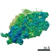
| Images | 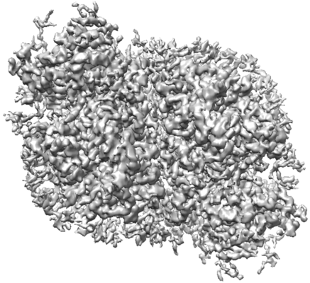 emd_30511.png emd_30511.png | 106.6 KB | ||
| Filedesc metadata | emd-30511.cif.gz | 8.6 KB | ||
| Archive directory |  http://ftp.pdbj.org/pub/emdb/structures/EMD-30511ftp://ftp.pdbj.org/pub/emdb/structures/EMD-30511 http://ftp.pdbj.org/pub/emdb/structures/EMD-30511ftp://ftp.pdbj.org/pub/emdb/structures/EMD-30511 | HTTPS FTP |
-Related structure data
| Related structure data |  7czlMC M: atomic model generated by this map C: citing same article ( |
|---|---|
| Similar structure data |

-Links
| EMDB pages | EMDB (EBI/PDBe) / EMDataResource |
|---|---|
| Related items in Molecule of the Month |


-Map
| File | Download / File: emd_30511.map.gz / Format: CCP4 / Size: 52.7 MB / Type: IMAGE STORED AS FLOATING POINT NUMBER (4 BYTES) | ||||||||||||||||||||||||||||||||||||||||||||||||||||||||||||
|---|---|---|---|---|---|---|---|---|---|---|---|---|---|---|---|---|---|---|---|---|---|---|---|---|---|---|---|---|---|---|---|---|---|---|---|---|---|---|---|---|---|---|---|---|---|---|---|---|---|---|---|---|---|---|---|---|---|---|---|---|---|
| Annotation | PSII-Psb27 | ||||||||||||||||||||||||||||||||||||||||||||||||||||||||||||
| Projections & slices | Image control
Images are generated by Spider. | ||||||||||||||||||||||||||||||||||||||||||||||||||||||||||||
| Voxel size | X=Y=Z: 1.30654 Å | ||||||||||||||||||||||||||||||||||||||||||||||||||||||||||||
| Density |
| ||||||||||||||||||||||||||||||||||||||||||||||||||||||||||||
| Symmetry | Space group: 1 | ||||||||||||||||||||||||||||||||||||||||||||||||||||||||||||
| Details | EMDB XML:
CCP4 map header:
| ||||||||||||||||||||||||||||||||||||||||||||||||||||||||||||
 Z (Sec.)
 Y (Row.)
 X (Col.)


-Supplemental data
- Sample components
+Entire : PSII-Psb27
+Supramolecule #1: PSII-Psb27
+Macromolecule #1: Photosystem II protein D1
+Macromolecule #2: Photosystem II CP47 reaction center protein
+Macromolecule #3: Photosystem II CP43 reaction center protein
+Macromolecule #4: Photosystem II D2 protein
+Macromolecule #5: Cytochrome b559 subunit alpha
+Macromolecule #6: Cytochrome b559 subunit beta
+Macromolecule #7: Photosystem II reaction center protein H
+Macromolecule #8: Photosystem II reaction center protein I
+Macromolecule #9: Photosystem II reaction center protein K
+Macromolecule #10: Photosystem II reaction center protein L
+Macromolecule #11: Photosystem II reaction center protein M
+Macromolecule #12: Photosystem II reaction center protein T
+Macromolecule #13: Photosystem II reaction center protein Z
+Macromolecule #14: Photosystem II reaction center protein Ycf12
+Macromolecule #15: Psb27
+Macromolecule #16: Photosystem II reaction center protein X
+Macromolecule #17: CHLOROPHYLL A

+Macromolecule #18: PHEOPHYTIN A

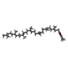
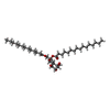
+Macromolecule #19: 5-[(2E,6E,10E,14E,18E,22E)-3,7,11,15,19,23,27-HEPTAMETHYLOCTACOSA...
+Macromolecule #20: BETA-CAROTENE

+Macromolecule #21: 1,2-DIPALMITOYL-PHOSPHATIDYL-GLYCEROLE

+Macromolecule #22: DODECYL-BETA-D-MALTOSIDE

+Macromolecule #23: 1,2-DI-O-ACYL-3-O-[6-DEOXY-6-SULFO-ALPHA-D-GLUCOPYRANOSYL]-SN-GLYCEROL

+Macromolecule #24: BICARBONATE ION

+Macromolecule #25: (1S)-2-(ALPHA-L-ALLOPYRANOSYLOXY)-1-[(TRIDECANOYLOXY)METHYL]ETHYL...
+Macromolecule #26: DIGALACTOSYL DIACYL GLYCEROL (DGDG)

+Macromolecule #27: CALCIUM ION
+Macromolecule #28: FE (II) ION
+Macromolecule #29: PROTOPORPHYRIN IX CONTAINING FE

+Macromolecule #30: CHLORIDE ION
-Experimental details
-Structure determination
| Method | cryo EM |
|---|---|
 Processing Processing | single particle reconstruction |
| Aggregation state | particle |
-Sample preparation
| Buffer | pH: 6 |
|---|---|
| Vitrification | Cryogen name: ETHANE |
- Electron microscopy
| Microscope | FEI TITAN KRIOS |
|---|---|
| Image recording | Film or detector model: GATAN K2 SUMMIT (4k x 4k) / Average electron dose: 50.0 e/Å2 |
| Electron beam | Acceleration voltage: 300 kV / Electron source:  FIELD EMISSION GUN FIELD EMISSION GUN |
| Electron optics | Illumination mode: FLOOD BEAM / Imaging mode: BRIGHT FIELD |
| Experimental equipment |  Model: Titan Krios / Image courtesy: FEI Company |
