| 登録情報 | データベース: PDB / ID: 4fuf
|
|---|
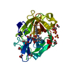
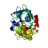
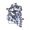
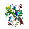
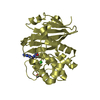
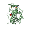
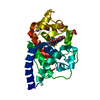
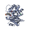
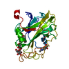
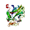
| タイトル | Crystal Structure of the Urokinase |
|---|
 要素 要素 | Urokinase-type plasminogen activator |
|---|
 キーワード キーワード | HYDROLASE/HYDROLASE INHIBITOR / HYDROLASE / HYDROLASE-HYDROLASE INHIBITOR complex |
|---|
| 機能・相同性 |  機能・相同性情報 機能・相同性情報
u-plasminogen activator / regulation of smooth muscle cell-matrix adhesion / urokinase plasminogen activator signaling pathway / regulation of plasminogen activation / regulation of fibrinolysis / regulation of wound healing / protein complex involved in cell-matrix adhesion / negative regulation of plasminogen activation / regulation of smooth muscle cell migration / regulation of signaling receptor activity ...u-plasminogen activator / regulation of smooth muscle cell-matrix adhesion / urokinase plasminogen activator signaling pathway / regulation of plasminogen activation / regulation of fibrinolysis / regulation of wound healing / protein complex involved in cell-matrix adhesion / negative regulation of plasminogen activation / regulation of smooth muscle cell migration / regulation of signaling receptor activity / serine-type endopeptidase complex / Dissolution of Fibrin Clot / smooth muscle cell migration / plasminogen activation / regulation of cell adhesion mediated by integrin / tertiary granule membrane / negative regulation of fibrinolysis / regulation of cell adhesion / specific granule membrane / serine protease inhibitor complex / fibrinolysis / chemotaxis / blood coagulation / regulation of cell population proliferation / response to hypoxia / positive regulation of cell migration / external side of plasma membrane / serine-type endopeptidase activity / focal adhesion / Neutrophil degranulation / cell surface / signal transduction / proteolysis / extracellular space / extracellular exosome / extracellular region / plasma membrane類似検索 - 分子機能 Kringle domain / Kringle / Kringle, conserved site / Kringle superfamily / Kringle domain signature. / Kringle domain profile. / Kringle domain / : / Kringle-like fold / EGF-like domain profile. ...Kringle domain / Kringle / Kringle, conserved site / Kringle superfamily / Kringle domain signature. / Kringle domain profile. / Kringle domain / : / Kringle-like fold / EGF-like domain profile. / EGF-like domain signature 1. / EGF-like domain / Serine proteases, trypsin family, histidine active site / Serine proteases, trypsin family, serine active site / Peptidase S1A, chymotrypsin family / Serine proteases, trypsin family, histidine active site. / Serine proteases, trypsin domain profile. / Serine proteases, trypsin family, serine active site. / Trypsin-like serine protease / Serine proteases, trypsin domain / Trypsin / Trypsin-like serine proteases / Thrombin, subunit H / Peptidase S1, PA clan, chymotrypsin-like fold / Peptidase S1, PA clan / Beta Barrel / Mainly Beta類似検索 - ドメイン・相同性 Chem-8UP / ACETATE ION / SUCCINIC ACID / Urokinase-type plasminogen activator類似検索 - 構成要素 |
|---|
| 生物種 |  Homo sapiens (ヒト) Homo sapiens (ヒト) |
|---|
| 手法 |  X線回折 / 分子置換 / 解像度: 2 Å X線回折 / 分子置換 / 解像度: 2 Å |
|---|
 データ登録者 データ登録者 | Kang, Y.N. / Stuckey, J.A. / Nienaber, V. / Giranda, V. |
|---|
 引用 引用 | ジャーナル: to be published
タイトル: Crystal Structure of the Urokinase
著者: Kang, Y.N. / Stuckey, J.A. / Nienaber, V. / Giranda, V. |
|---|
| 履歴 | | 登録 | 2012年6月28日 | 登録サイト: RCSB / 処理サイト: RCSB |
|---|
| 改定 1.0 | 2012年8月22日 | Provider: repository / タイプ: Initial release |
|---|
| 改定 1.1 | 2017年11月15日 | Group: Refinement description / カテゴリ: software
Item: _software.classification / _software.contact_author ..._software.classification / _software.contact_author / _software.contact_author_email / _software.date / _software.language / _software.location / _software.name / _software.type / _software.version |
|---|
|
|---|
 ムービー
ムービー コントローラー


 データを開く
 基本情報
基本情報 構造の表示
構造の表示 ダウンロードとリンク
 その他のダウンロード


 PDBj
PDBj

 集合体
集合体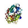


 分子量: 337.212 Da / 分子数: 1 / 由来タイプ: 合成 / 式: C15H17BrN2O2
 分子量: 118.088 Da / 分子数: 1 / 由来タイプ: 合成 / 式: C4H6O4
 分子量: 96.063 Da / 分子数: 3 / 由来タイプ: 合成 / 式: SO4
 分子量: 92.094 Da / 分子数: 4 / 由来タイプ: 合成 / 式: C3H8O3
 分子量: 59.044 Da / 分子数: 3 / 由来タイプ: 合成 / 式: C2H3O2
 試料調製
試料調製 解析
解析