Mass: 18.015 Da / Num. of mol.: 6 / Source method: isolated from a natural source / Formula: H2O
-
Experimental details
-
Experiment
Experiment
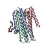
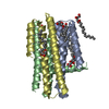
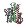
Method: X-RAY DIFFRACTION / Number of used crystals: 1
-
Sample preparation
Crystal
Density Matthews: 3.9 Å3/Da / Density % sol: 68.47 % / Description: NONE
Crystal grow
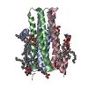
Temperature: 277 K / Method: lipidic cubic phase / pH: 5.6 Details: 4-6 %(V/V) 2-METHYL-2, 4-PENTANEDIOL (MPD), 0.1 M SODIUM CHLORIDE, 60 MM MAGNESIUM ACETATE, 0.1 M SODIUM CITRATE/HCL PH 5.6. CRYSTALLIZED USING THE IN MESO (LIPIDIC CUBIC PHASE) METHOD AT 4 ...Details: 4-6 %(V/V) 2-METHYL-2, 4-PENTANEDIOL (MPD), 0.1 M SODIUM CHLORIDE, 60 MM MAGNESIUM ACETATE, 0.1 M SODIUM CITRATE/HCL PH 5.6. CRYSTALLIZED USING THE IN MESO (LIPIDIC CUBIC PHASE) METHOD AT 4 DEGREES CELCIUS WITH THE 7.9 MONOACYLGLYCEROL (7.9 MAG) AS THE HOSTING LIPID.
Resolution: 2.44→66.533 Å / SU ML: 0.32 / σ(F): 1.36 / Phase error: 28.12 / Stereochemistry target values: ML Details: THERE ARE THREE NCS-RELATED MOLECULES IN THE ASYMMETRIC UNIT BUT THE NCS RESTRAINTS WERE NOT USED IN THE REFINEMENT.
Rfactor
Num. reflection
% reflection
Rfree
0.2522
1847
5 %
Rwork
0.2178
-
-
obs
0.2195
37090
98.71 %
Solvent computation
Shrinkage radii: 0.9 Å / VDW probe radii: 1.11 Å / Solvent model: FLAT BULK SOLVENT MODEL
Displacement parameters
Biso mean: 73.27 Å2
Refinement step
Cycle: LAST / Resolution: 2.44→66.533 Å
Protein
Nucleic acid
Ligand
Solvent
Total
Num. atoms
4470
0
87
6
4563
Refine LS restraints
Refine-ID
Type
Dev ideal
Number
X-RAY DIFFRACTION
f_bond_d
0.007
4627
X-RAY DIFFRACTION
f_angle_d
0.688
6307
X-RAY DIFFRACTION
f_dihedral_angle_d
11.428
1603
X-RAY DIFFRACTION
f_chiral_restr
0.045
798
X-RAY DIFFRACTION
f_plane_restr
0.002
751
LS refinement shell
Resolution (Å)
Rfactor Rfree
Num. reflection Rfree
Rfactor Rwork
Num. reflection Rwork
Refine-ID
% reflection obs (%)
2.44-2.506
0.3586
133
0.3257
2692
X-RAY DIFFRACTION
99
2.506-2.5797
0.3466
121
0.2838
2704
X-RAY DIFFRACTION
99
2.5797-2.663
0.2813
157
0.256
2671
X-RAY DIFFRACTION
99
2.663-2.7582
0.2966
131
0.2442
2672
X-RAY DIFFRACTION
99
2.7582-2.8686
0.2614
131
0.228
2707
X-RAY DIFFRACTION
99
2.8686-2.9992
0.2596
147
0.2163
2670
X-RAY DIFFRACTION
98
2.9992-3.1573
0.2353
150
0.2076
2687
X-RAY DIFFRACTION
99
3.1573-3.3551
0.2358
149
0.2142
2698
X-RAY DIFFRACTION
99
3.3551-3.6141
0.2532
147
0.206
2711
X-RAY DIFFRACTION
99
3.6141-3.9778
0.2303
146
0.1971
2711
X-RAY DIFFRACTION
98
3.9778-4.5533
0.222
152
0.1741
2728
X-RAY DIFFRACTION
99
4.5533-5.7361
0.2788
137
0.2242
2754
X-RAY DIFFRACTION
98
5.7361-66.5574
0.2423
146
0.2321
2838
X-RAY DIFFRACTION
97
Refinement TLS params.
Method: refined / Refine-ID: X-RAY DIFFRACTION
ID
L11 (°2)
L12 (°2)
L13 (°2)
L22 (°2)
L23 (°2)
L33 (°2)
S11 (Å °)
S12 (Å °)
S13 (Å °)
S21 (Å °)
S22 (Å °)
S23 (Å °)
S31 (Å °)
S32 (Å °)
S33 (Å °)
T11 (Å2)
T12 (Å2)
T13 (Å2)
T22 (Å2)
T23 (Å2)
T33 (Å2)
Origin x (Å)
Origin y (Å)
Origin z (Å)
1
7.9694
-5.6009
-2.4101
4.9035
0.8812
4.9972
-0.9183
-0.7912
0.6814
0.8911
0.7454
-0.3926
-0.9082
-0.4823
0.1817
0.621
0.027
-0.0435
0.7282
-0.1661
0.3962
-1.2586
-11.5397
22.5959
2
4.7179
-0.692
0.3414
1.7592
1.7454
1.9708
-0.1985
-0.0584
0.0545
0.1268
0.026
-0.1986
0.2974
-0.2336
0.1873
0.286
0.0063
0.0136
0.3561
0.0084
0.306
-1.0176
-21.3543
9.4533
3
7.9221
1.347
-1.8314
6.9886
-0.2235
0.4394
0.216
-0.1245
-0.0809
-0.0318
-0.1082
-0.0835
0.2235
-0.2347
0.1424
0.33
0.0587
-0.0382
0.2504
-0.1046
0.3812
-6.2389
-31.3102
9.7274
4
8.6312
-1.7901
0.9789
9.0562
1.2111
7.3647
-0.3585
-1.1625
0.2571
1.0916
0.4546
0.6849
-1.1619
-0.64
-0.1603
0.4277
0.1345
0.0048
0.5447
0.0253
0.6381
-21.7865
-7.4649
8.8024
5
3.8534
1.0415
-0.0557
3.107
-1.5188
5.9044
0.0019
-0.1303
0.1659
0.4412
0.16
0.0568
-0.8305
-0.049
-0.1585
0.3441
0.0566
0.0138
0.2212
-0.0286
0.315
-10.8741
-14.4062
13.8918
6
7.6102
3.9816
-2.7496
3.6614
-4.0462
6.6209
0.4234
0.7768
-1.2747
-0.7271
-0.7924
-0.339
-0.0493
0.0823
0.6246
0.8311
0.0785
-0.1935
0.4939
-0.1899
0.7983
-19.5387
-33.5442
-4.427
7
3.5363
-0.6793
1.416
2.5949
-0.2975
8.4735
0.1404
-0.4389
-0.0979
0.3312
0.0796
0.3208
0.3064
-0.6576
-0.1715
0.321
-0.0749
0.0099
0.4034
0.0525
0.3159
-20.0418
-24.4071
10.5227
8
2.9596
0.8344
1.4348
5.3467
-0.4116
3.721
-0.0846
0.0846
-0.1321
0.1009
0.0752
-0.0306
-0.1418
0.0353
0.0227
0.3382
0.0676
0.0233
0.4002
0.039
0.3302
-33.9663
-3.9623
-9.657
9
5.5481
0.3168
-1.222
5.2216
-4.0916
8.6125
-0.256
0.1179
-0.0163
0.0804
-0.3489
-0.4164
-0.3802
0.9316
0.6132
0.3196
0.0048
-0.0914
0.3771
0.0023
0.4905
-24.2008
3.8292
-9.9552
10
1.4976
0.5843
0.6628
0.9155
0.4804
0.5339
-0.6892
-0.9435
1.2481
0.4251
0.0945
0.6099
0.0802
-0.3414
0.515
0.5624
0.6589
0.3005
1.6939
0.3796
1.7086
-50.3845
13.922
0.8099
11
3.4498
1.3322
1.0609
2.9263
-0.6317
1.7943
0.1062
0.1186
0.2117
0.0198
0.0483
0.1002
-0.5201
-1.2106
-0.1548
0.392
0.1493
0.0133
0.4784
0.0637
0.3774
-42.3759
5.0068
-12.5078
12
2.5344
3.0404
-3.0456
3.6499
-3.6554
3.6635
-0.5137
-0.9797
-0.1689
0.379
-0.5882
-1.4067
-1.4428
0.3289
0.9038
1.3318
-0.134
-0.1348
1.0664
0.0124
1.1224
-22.5599
18.5245
0.9444
13
2.1316
-0.8374
-0.2204
2.9141
-2.4799
7.9245
0.2053
0.0706
-0.1483
-0.0295
0.0893
-0.0534
-1.1432
0.6098
-0.3731
0.421
-0.0381
0.0196
0.369
-0.0204
0.4442
-30.5145
11.6421
-17.5436
14
6.9903
1.3312
0.9919
5.0012
3.3405
3.7187
-0.2001
-0.6951
0.5778
0.2155
-0.0262
-0.0461
-2.3101
-1.0236
0.3283
0.8563
0.2025
-0.0697
0.3755
0.0117
0.4824
-36.661
19.2626
-5.9159
Refinement TLS group
ID
Refine-ID
Refine TLS-ID
Selection details
1
X-RAY DIFFRACTION
1
CHAINAAND (RESID8THROUGH27 )
2
X-RAY DIFFRACTION
2
CHAINAAND (RESID28THROUGH82 )
3
X-RAY DIFFRACTION
3
CHAINAAND (RESID83THROUGH121 )
4
X-RAY DIFFRACTION
4
CHAINBAND (RESID23THROUGH51 )
5
X-RAY DIFFRACTION
5
CHAINBAND (RESID52THROUGH120 )
6
X-RAY DIFFRACTION
6
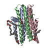
CHAINCAND (RESID33THROUGH51 )
7
X-RAY DIFFRACTION
7
CHAINCAND (RESID52THROUGH121 )
8
X-RAY DIFFRACTION
8
CHAINDAND (RESID14THROUGH82 )
9
X-RAY DIFFRACTION
9
CHAINDAND (RESID83THROUGH121 )
10
X-RAY DIFFRACTION
10
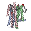
CHAINEAND (RESID34THROUGH51 )
11
X-RAY DIFFRACTION
11
CHAINEAND (RESID52THROUGH120 )
12
X-RAY DIFFRACTION
12
CHAINFAND (RESID33THROUGH51 )
13
X-RAY DIFFRACTION
13
CHAINFAND (RESID52THROUGH90 )
14
X-RAY DIFFRACTION
14
CHAINFAND (RESID91THROUGH120 )
+
About Yorodumi
-
News
-
Feb 9, 2022. New format data for meta-information of EMDB entries
New format data for meta-information of EMDB entries
Version 3 of the EMDB header file is now the official format.
The previous official version 1.9 will be removed from the archive.
In the structure databanks used in Yorodumi, some data are registered as the other names, "COVID-19 virus" and "2019-nCoV". Here are the details of the virus and the list of structure data.
Jan 31, 2019. EMDB accession codes are about to change! (news from PDBe EMDB page)
EMDB accession codes are about to change! (news from PDBe EMDB page)
The allocation of 4 digits for EMDB accession codes will soon come to an end. Whilst these codes will remain in use, new EMDB accession codes will include an additional digit and will expand incrementally as the available range of codes is exhausted. The current 4-digit format prefixed with “EMD-” (i.e. EMD-XXXX) will advance to a 5-digit format (i.e. EMD-XXXXX), and so on. It is currently estimated that the 4-digit codes will be depleted around Spring 2019, at which point the 5-digit format will come into force.
The EM Navigator/Yorodumi systems omit the EMD- prefix.
Related info.:Q: What is EMD? / ID/Accession-code notation in Yorodumi/EM Navigator
Yorodumi is a browser for structure data from EMDB, PDB, SASBDB, etc.
This page is also the successor to EM Navigator detail page, and also detail information page/front-end page for Omokage search.
The word "yorodu" (or yorozu) is an old Japanese word meaning "ten thousand". "mi" (miru) is to see.
Related info.:EMDB / PDB / SASBDB / Comparison of 3 databanks / Yorodumi Search / Aug 31, 2016. New EM Navigator & Yorodumi / Yorodumi Papers / Jmol/JSmol / Function and homology information / Changes in new EM Navigator and Yorodumi
 Movie
Movie Controller

 Yorodumi
 Open data
 Basic information
 Components
 Keywords
 Function and homology information
 X-RAY DIFFRACTION /
  Authors
 Citation
 Structure visualization
 Downloads & links
 Other downloads
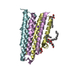


 PDBj
PDBj


 Assembly
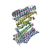


 Mass: 328.487 Da / Num. of mol.: 3 / Source method: obtained synthetically / Formula: C19H36O4
 Mass: 65.409 Da / Num. of mol.: 1 / Source method: obtained synthetically / Formula: Zn
 Mass: 59.044 Da / Num. of mol.: 1 / Source method: obtained synthetically / Formula: C2H3O2
 Mass: 189.100 Da / Num. of mol.: 1 / Source method: obtained synthetically / Formula: C6H5O7
 Sample preparation
 / Beamline: X06DA / Wavelength: 1.03315
  Processing