[English] 日本語
Yorodumi
- EMDB-4100: Structure of F-ATPase from Pichia angusta, state3 -

+
Open data


ID or keywords:

Loading...

-
Basic information

Entry
Database: EMDB / ID: EMD-4100
TitleStructure of F-ATPase from Pichia angusta, state3
Map data
Sample
  • Complex: Yeast F1FO ATP Synthase
    • Protein or peptide: x 17 types
  • Ligand: x 3 types
KeywordsATP synthase / hydrolase
Function / homology
Function and homology information


negative regulation of mitochondrial ATP synthesis coupled proton transport / angiostatin binding / ATPase inhibitor activity / negative regulation of hydrolase activity / : / : / heme biosynthetic process / : / : / : ...negative regulation of mitochondrial ATP synthesis coupled proton transport / angiostatin binding / ATPase inhibitor activity / negative regulation of hydrolase activity / : / : / heme biosynthetic process / : / : / : / proton-transporting ATP synthase complex, coupling factor F(o) / negative regulation of endothelial cell proliferation / proton motive force-driven ATP synthesis / proton motive force-driven mitochondrial ATP synthesis / proton transmembrane transporter activity / proton-transporting ATP synthase complex, catalytic core F(1) / H+-transporting two-sector ATPase / proton-transporting ATPase activity, rotational mechanism / proton-transporting ATP synthase activity, rotational mechanism / erythrocyte differentiation / ADP binding / ATPase binding / protein homotetramerization / mitochondrial inner membrane / calmodulin binding / lipid binding / structural molecule activity / cell surface / protein homodimerization activity / ATP hydrolysis activity / protein-containing complex / mitochondrion / ATP binding / identical protein binding
Similarity search - Function
: / Fungal epsilon subunit of F1F0-ATP synthase C-terminal domain / Mitochondrial ATPase inhibitor / Mitochondrial ATPase inhibitor, IATP / ATP synthase, F0 complex, subunit B/MI25 / ATP synthase, F0 complex, subunit B / Mitochondrial ATP synthase B chain precursor (ATP-synt_B) / ATP synthase, F0 complex, subunit D, mitochondrial / ATP synthase D chain, mitochondrial (ATP5H) / ATP synthase, F0 complex, subunit D superfamily, mitochondrial ...: / Fungal epsilon subunit of F1F0-ATP synthase C-terminal domain / Mitochondrial ATPase inhibitor / Mitochondrial ATPase inhibitor, IATP / ATP synthase, F0 complex, subunit B/MI25 / ATP synthase, F0 complex, subunit B / Mitochondrial ATP synthase B chain precursor (ATP-synt_B) / ATP synthase, F0 complex, subunit D, mitochondrial / ATP synthase D chain, mitochondrial (ATP5H) / ATP synthase, F0 complex, subunit D superfamily, mitochondrial / ATP synthase, F0 complex, subunit A, bacterial/mitochondria / ATP synthase, F1 complex, epsilon subunit, mitochondrial / ATP synthase, F1 complex, epsilon subunit superfamily, mitochondrial / Mitochondrial ATP synthase epsilon chain / F1F0 ATP synthase OSCP/delta subunit, N-terminal domain superfamily / ATP synthase, F0 complex, subunit A / ATP synthase, F0 complex, subunit A, active site / ATP synthase, F0 complex, subunit A superfamily / ATP synthase A chain / ATP synthase a subunit signature. / ATPase, OSCP/delta subunit / ATP synthase delta (OSCP) subunit / ATP synthase, F1 complex, delta/epsilon subunit / ATP synthase, F1 complex, delta/epsilon subunit, N-terminal / F0F1 ATP synthase delta/epsilon subunit, N-terminal / ATP synthase, Delta/Epsilon chain, beta-sandwich domain / ATP synthase, F0 complex, subunit C / F1F0 ATP synthase subunit C superfamily / ATP synthase, F0 complex, subunit C, DCCD-binding site / ATP synthase c subunit signature. / ATP synthase, F1 complex, gamma subunit conserved site / ATP synthase gamma subunit signature. / ATP synthase, F1 complex, beta subunit / ATP synthase, alpha subunit, C-terminal domain superfamily / : / ATP synthase, F1 complex, gamma subunit / ATP synthase, F1 complex, gamma subunit superfamily / ATP synthase / ATP synthase, alpha subunit, C-terminal / ATP synthase, F1 complex, alpha subunit / ATP synthase, F1 complex, alpha subunit nucleotide-binding domain / ATP synthase alpha/beta chain, C terminal domain / V-ATPase proteolipid subunit C-like domain / F/V-ATP synthase subunit C superfamily / ATP synthase subunit C / ATPase, F1/V1 complex, beta/alpha subunit, C-terminal / C-terminal domain of V and A type ATP synthase / ATP synthase subunit alpha, N-terminal domain-like superfamily / ATPase, F1/V1/A1 complex, alpha/beta subunit, N-terminal domain superfamily / ATPase, F1/V1/A1 complex, alpha/beta subunit, N-terminal domain / ATP synthase alpha/beta family, beta-barrel domain / ATPase, alpha/beta subunit, nucleotide-binding domain, active site / ATP synthase alpha and beta subunits signature. / ATPase, F1/V1/A1 complex, alpha/beta subunit, nucleotide-binding domain / ATP synthase alpha/beta family, nucleotide-binding domain / ATPases associated with a variety of cellular activities / AAA+ ATPase domain / P-loop containing nucleoside triphosphate hydrolase
Similarity search - Domain/homology
ATP synthase subunit epsilon, mitochondrial / ATP synthase subunit alpha, mitochondrial / ATP synthase subunit beta, mitochondrial / ATP synthase subunit gamma, mitochondrial / ATP synthase subunit 4, mitochondrial / ATP synthase subunit 9, mitochondrial / ATP synthase subunit d, mitochondrial / ATP synthase subunit a / ATPase inhibitor, mitochondrial / ATP synthase subunit delta, mitochondrial / ATP synthase subunit 5, mitochondrial
Similarity search - Component
Biological speciesOgataea angusta (fungus) / Bos taurus (cattle)
Methodsingle particle reconstruction / cryo EM / Resolution: 7.9 Å
AuthorsVinothkumar KR / Montgomery MG
CitationJournal: Proc Natl Acad Sci U S A / Year: 2016
Title: Structure of the mitochondrial ATP synthase from determined by electron cryo-microscopy.
Authors: Kutti R Vinothkumar / Martin G Montgomery / Sidong Liu / John E Walker /
Abstract: The structure of the intact monomeric ATP synthase from the fungus, , has been solved by electron cryo-microscopy. The structure provides insights into the mechanical coupling of the transmembrane ...The structure of the intact monomeric ATP synthase from the fungus, , has been solved by electron cryo-microscopy. The structure provides insights into the mechanical coupling of the transmembrane proton motive force across mitochondrial membranes in the synthesis of ATP. This mechanism requires a strong and integral stator, consisting of the catalytic αβ-domain, peripheral stalk, and, in the membrane domain, subunit a and associated supernumerary subunits, kept in contact with the rotor turning at speeds up to 350 Hz. The stator's integrity is ensured by robust attachment of both the oligomycin sensitivity conferral protein (OSCP) to the catalytic domain and the membrane domain of subunit b to subunit a. The ATP8 subunit provides an additional brace between the peripheral stalk and subunit a. At the junction between the OSCP and the apparently stiff, elongated α-helical b-subunit and associated d- and h-subunits, an elbow or joint allows the stator to bend to accommodate lateral movements during the activity of the catalytic domain. The stator may also apply lateral force to help keep the static a-subunit and rotating c-ring together. The interface between the c-ring and the a-subunit contains the transmembrane pathway for protons, and their passage across the membrane generates the turning of the rotor. The pathway has two half-channels containing conserved polar residues provided by a bundle of four α-helices inclined at ∼30° to the plane of the membrane, similar to those described in other species. The structure provides more insights into the workings of this amazing machine.
History
DepositionOct 4, 2016-
Header (metadata) releaseNov 16, 2016-
Map releaseNov 16, 2016-
UpdateMay 15, 2024-
Current statusMay 15, 2024Processing site: PDBe / Status: Released

-
Structure visualization

Movie
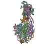
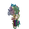
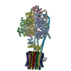
  • Surface view with section colored by density value
  • Surface level: 0.15
  • Imaged by UCSF Chimera
  • Download
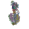
  • Surface view colored by cylindrical radius
  • Surface level: 0.15
  • Imaged by UCSF Chimera
  • Download
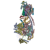
  • Surface view with fitted model
  • Atomic models: PDB-5lqx
  • Surface level: 0.15
  • Imaged by UCSF Chimera
  • Download
Movie viewer
Structure viewerEM map:
SurfViewMolmilJmol/JSmol
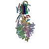
Supplemental images

Downloads & links

-
Map

FileDownload / File: emd_4100.map.gz / Format: CCP4 / Size: 103 MB / Type: IMAGE STORED AS FLOATING POINT NUMBER (4 BYTES)
Projections & slices

Image control

Size
Brightness
Contrast
Others
AxesZ (Sec.)Y (Row.)X (Col.)
1.75 Å/pix.
x 300 pix.
= 525. Å
1.75 Å/pix.
x 300 pix.
= 525. Å
1.75 Å/pix.
x 300 pix.
= 525. Å

Surface

Projections

Slices (1/3)

Slices (1/2)

Slices (2/3)

Images are generated by Spider.

Voxel sizeX=Y=Z: 1.75 Å
Density
Contour LevelBy AUTHOR: 0.1 / Movie #1: 0.15
Minimum - Maximum-0.16187143 - 0.51867765
Average (Standard dev.)0.00009730653 (±0.017747326)
SymmetrySpace group: 1
Details

EMDB XML:

Map geometry
Axis orderXYZ
Origin000
Dimensions300300300
Spacing300300300
CellA=B=C: 525.0 Å
α=β=γ: 90.0 °

CCP4 map header:

modeImage stored as Reals
Å/pix. X/Y/Z1.751.751.75
M x/y/z300300300
origin x/y/z0.0000.0000.000
length x/y/z525.000525.000525.000
α/β/γ90.00090.00090.000
MAP C/R/S123
start NC/NR/NS000
NC/NR/NS300300300
D min/max/mean-0.1620.5190.000

-
Supplemental data

-
Sample components

+
Entire : Yeast F1FO ATP Synthase

EntireName: Yeast F1FO ATP Synthase
Components
  • Complex: Yeast F1FO ATP Synthase
    • Protein or peptide: ATP synthase subunit f
    • Protein or peptide: ATP synthase AAP1 subunit
    • Protein or peptide: ATP synthase subunit a
    • Protein or peptide: ATP synthase subunit b
    • Protein or peptide: ATP synthase alpha subunit
    • Protein or peptide: ATP synthase beta subunit
    • Protein or peptide: ATP synthase gamma subunit
    • Protein or peptide: ATP synthase delta subunit
    • Protein or peptide: ATP synthase epsilon subunit
    • Protein or peptide: ATP synthase inhibitor protein IF1
    • Protein or peptide: ATP synthase subunit c
    • Protein or peptide: ATP synthase OSCP subunit
    • Protein or peptide: ATP synthase subunit b
    • Protein or peptide: ATP synthase subunit d
    • Protein or peptide: ATP synthase subunit h
    • Protein or peptide: ATP synthase subunit a
    • Protein or peptide: ATP synthase subunit a
  • Ligand: ADENOSINE-5'-TRIPHOSPHATE
  • Ligand: MAGNESIUM ION
  • Ligand: ADENOSINE-5'-DIPHOSPHATE

+
Supramolecule #1: Yeast F1FO ATP Synthase

SupramoleculeName: Yeast F1FO ATP Synthase / type: complex / ID: 1 / Parent: 0 / Macromolecule list: #1-#17
Details: The complex comprises of the catalytic domain, membrane domain and peripheral stalk of the yeast ATP synthase.
Source (natural)Organism: Ogataea angusta (fungus) / Strain: A16
Molecular weightTheoretical: 550 KDa

+
Macromolecule #1: ATP synthase subunit f

MacromoleculeName: ATP synthase subunit f / type: protein_or_peptide / ID: 1 / Number of copies: 1 / Enantiomer: LEVO
Source (natural)Organism: Ogataea angusta (fungus)
Molecular weightTheoretical: 2.656265 KDa
SequenceString:
(UNK)(UNK)(UNK)(UNK)(UNK)(UNK)(UNK)(UNK)(UNK)(UNK) (UNK)(UNK)(UNK)(UNK)(UNK)(UNK) (UNK)(UNK)(UNK) (UNK)(UNK)(UNK)(UNK)(UNK)(UNK)(UNK)(UNK)(UNK)(UNK) (UNK)(UNK)

+
Macromolecule #2: ATP synthase AAP1 subunit

MacromoleculeName: ATP synthase AAP1 subunit / type: protein_or_peptide / ID: 2 / Number of copies: 1 / Enantiomer: LEVO
Source (natural)Organism: Ogataea angusta (fungus)
Molecular weightTheoretical: 2.145636 KDa
SequenceString:
(UNK)(UNK)(UNK)(UNK)(UNK)(UNK)(UNK)(UNK)(UNK)(UNK) (UNK)(UNK)(UNK)(UNK)(UNK)(UNK) (UNK)(UNK)(UNK) (UNK)(UNK)(UNK)(UNK)(UNK)(UNK)

+
Macromolecule #3: ATP synthase subunit a

MacromoleculeName: ATP synthase subunit a / type: protein_or_peptide / ID: 3 / Details: Helix 1 of subunit a / Number of copies: 1 / Enantiomer: LEVO
Source (natural)Organism: Ogataea angusta (fungus)
Molecular weightTheoretical: 1.464797 KDa
SequenceString:
(UNK)(UNK)(UNK)(UNK)(UNK)(UNK)(UNK)(UNK)(UNK)(UNK) (UNK)(UNK)(UNK)(UNK)(UNK)(UNK) (UNK)

+
Macromolecule #4: ATP synthase subunit b

MacromoleculeName: ATP synthase subunit b / type: protein_or_peptide / ID: 4 / Details: Helix 1 of subunit b / Number of copies: 1 / Enantiomer: LEVO
Source (natural)Organism: Ogataea angusta (fungus)
Molecular weightTheoretical: 2.315846 KDa
SequenceString:
(UNK)(UNK)(UNK)(UNK)(UNK)(UNK)(UNK)(UNK)(UNK)(UNK) (UNK)(UNK)(UNK)(UNK)(UNK)(UNK) (UNK)(UNK)(UNK) (UNK)(UNK)(UNK)(UNK)(UNK)(UNK)(UNK)(UNK)

+
Macromolecule #5: ATP synthase alpha subunit

MacromoleculeName: ATP synthase alpha subunit / type: protein_or_peptide / ID: 5 / Number of copies: 3 / Enantiomer: LEVO
Source (natural)Organism: Ogataea angusta (fungus)
Molecular weightTheoretical: 55.007535 KDa
SequenceString: ATAKAAPTEV SSILESKIRG VSDEANLDET GRVLSVGDGI ARVFGLNNCQ AEELVEFASG VKGMALNLEP GQVGIVLFGS DREVKEGEI VKRTGKIVDV PIGPGMLGRV VDALGNPIDG KGPIEATGYA IAQLKAPGIL PRRSVFEPMQ TGLKAVDALV P IGRGQREL ...String:
ATAKAAPTEV SSILESKIRG VSDEANLDET GRVLSVGDGI ARVFGLNNCQ AEELVEFASG VKGMALNLEP GQVGIVLFGS DREVKEGEI VKRTGKIVDV PIGPGMLGRV VDALGNPIDG KGPIEATGYA IAQLKAPGIL PRRSVFEPMQ TGLKAVDALV P IGRGQREL IIGDRQTGKT AVALDTILNQ KRWNDGNDES KKLYCVYVAV GQKRSTVAQL VQTLEQNDAM KYSIVVAATA SE AAPLQYL APFTACAIAE WFRDNGKHAL IVYDDLSKQA VAYRQLSLLL RRPPGREAYP GDVFYLHSRL LERAAKMSDA NGG GSLTAL PVIETQGGDV SAYIPTNVIS ITDGQIFLEA ELFYKGIRPA INVGLSVSRV GSAAQVKAMK QVAGSLKLFL AQYR EVAAF AQFGSDLDAS TKQTLSRGER LTQLLKQKQY SPQASEEQVP VIYAGVNGFL DNIPIERIPE FEEQFIAYLK ANEGD ILEA IRTKGELSSE LLDKLKSATE TFVATF

+
Macromolecule #6: ATP synthase beta subunit

MacromoleculeName: ATP synthase beta subunit / type: protein_or_peptide / ID: 6 / Number of copies: 3 / Enantiomer: LEVO
Source (natural)Organism: Ogataea angusta (fungus)
Molecular weightTheoretical: 50.852434 KDa
SequenceString: ATAGPASGKI RAVIGAVVDV QFEQGELPAI LNALTIDQGN NQKLVLEVAQ HLGENAVRAI AMDGTEGLVR GQTVVDTGAP ISVPVGRGT LGRIINVVGE PIDERGPIEC KQRNPIHADP PSFVEQSTEA EVLETGIKVV DLLAPYARGG KIGLFGGAGV G KTVFIQEL ...String:
ATAGPASGKI RAVIGAVVDV QFEQGELPAI LNALTIDQGN NQKLVLEVAQ HLGENAVRAI AMDGTEGLVR GQTVVDTGAP ISVPVGRGT LGRIINVVGE PIDERGPIEC KQRNPIHADP PSFVEQSTEA EVLETGIKVV DLLAPYARGG KIGLFGGAGV G KTVFIQEL INNIAKAHGG FSVFTGVGER TREGNDLYRE MKETGVINLE GESKVALVFG QMNEPPGARA RVALTGLTIA EY FRDEEGQ DVLLFVDNIF RFTQAGSEVS ALLGRIPSAV GYQPTLATDM GLLQERITTT RKGSVTSVQA VYVPADDLTD PAP ATTFAH LDATTVLSRG ISELGIYPAV DPLDSKSRLL DVSVVGQEHY DVATGVQQTL QAYKSLQDII AILGMDELSE QDKL TVERA RKIQRFLSQP FAVAEVFTGI EGKLVRLKDT IASFKAVLEG KYDHLPENAF YMVGGIEDVV AKAEKIAAEA N

+
Macromolecule #7: ATP synthase gamma subunit

MacromoleculeName: ATP synthase gamma subunit / type: protein_or_peptide / ID: 7 / Number of copies: 1 / Enantiomer: LEVO
Source (natural)Organism: Ogataea angusta (fungus)
Molecular weightTheoretical: 29.418238 KDa
SequenceString: ATLREIETRL KSIKNIEKIT NTMKVVASTR MGRAQRAMAS SRAFREGDSD FFATAETSTP ETAEKTLIIA VSSDKGLCGS IHSQIAKAT RAKLQETPNA DVVTIGDKIK AQMLRTHSSN VVLSFNGVGK EAPTFWEASL IADEIRKLGD YDKIEVMYNK F VSGVAFEP ...String:
ATLREIETRL KSIKNIEKIT NTMKVVASTR MGRAQRAMAS SRAFREGDSD FFATAETSTP ETAEKTLIIA VSSDKGLCGS IHSQIAKAT RAKLQETPNA DVVTIGDKIK AQMLRTHSSN VVLSFNGVGK EAPTFWEASL IADEIRKLGD YDKIEVMYNK F VSGVAFEP SVFPSFSPIS IEESPKLSEF ELEEDQAIPT SLSQISLTNA ILNAMAEGYA SEISARRNAM DNASKNAGEM IN KYSILYN RTRQAVITNE LVDIITGASS LD

+
Macromolecule #8: ATP synthase delta subunit

MacromoleculeName: ATP synthase delta subunit / type: protein_or_peptide / ID: 8 / Number of copies: 1 / Enantiomer: LEVO
Source (natural)Organism: Ogataea angusta (fungus)
Molecular weightTheoretical: 14.171783 KDa
SequenceString:
AEAVNPDVLK VSLVAPHQAI FTNKEVSQVN LPASSGEMGV LANHVPTVEE LAPGVVEVIE SSGTASKYFV SGGFASILPG SKLSISTVE AHPLDAFSSE NIKSLLAEAQ KNASSADETV AAEAAIEIEV LEALQAAVH

+
Macromolecule #9: ATP synthase epsilon subunit

MacromoleculeName: ATP synthase epsilon subunit / type: protein_or_peptide / ID: 9 / Number of copies: 1 / Enantiomer: LEVO
Source (natural)Organism: Ogataea angusta (fungus)
Molecular weightTheoretical: 6.910886 KDa
SequenceString:
SSWQKAGISF NKYLAIAART VQRSLKNDLK VAAEKRYISD AKVQKLEKGN VVSTTDLASN KSA

+
Macromolecule #10: ATP synthase inhibitor protein IF1

MacromoleculeName: ATP synthase inhibitor protein IF1 / type: protein_or_peptide / ID: 10 / Number of copies: 1 / Enantiomer: LEVO
Source (natural)Organism: Bos taurus (cattle)
Molecular weightTheoretical: 7.462098 KDa
Recombinant expressionOrganism: Escherichia coli BL21(DE3) (bacteria)
SequenceString:
GSESGDNVRS SAGAVRDAGG AFGKREQAEE ERYFRARAKE QLAALKKHHE NEISHHAKEI HHHHHH

+
Macromolecule #11: ATP synthase subunit c

MacromoleculeName: ATP synthase subunit c / type: protein_or_peptide / ID: 11 / Number of copies: 10 / Enantiomer: LEVO
Source (natural)Organism: Ogataea angusta (fungus)
Molecular weightTheoretical: 7.83738 KDa
SequenceString:
MQLVLAAKYI GAAIATIGLT GAGIGIAIVF AALINGTSRN PSLRNTLFPF AILGFALSEA TGLFCLMISF LLLYGV

+
Macromolecule #12: ATP synthase OSCP subunit

MacromoleculeName: ATP synthase OSCP subunit / type: protein_or_peptide / ID: 12 / Number of copies: 1 / Enantiomer: LEVO
Source (natural)Organism: Ogataea angusta (fungus)
Molecular weightTheoretical: 20.678812 KDa
SequenceString: ASAAPIKPPV QLFGLDGTYA TALFSASAKD SSIEKTFQSV QKLSSTISKD AKVAQVLSNP ALSLNSRKEV VSVLSKELKL EPVVSNLLT VLAENNRLSL FDSIAKQFSV LNDAYNGVVE ATVVSAKPLD SKILNRLTKS ITNSKYVGPG KTLKIKNEVD P EILGGLIV ...String:
ASAAPIKPPV QLFGLDGTYA TALFSASAKD SSIEKTFQSV QKLSSTISKD AKVAQVLSNP ALSLNSRKEV VSVLSKELKL EPVVSNLLT VLAENNRLSL FDSIAKQFSV LNDAYNGVVE ATVVSAKPLD SKILNRLTKS ITNSKYVGPG KTLKIKNEVD P EILGGLIV EVADKSVDLS LASKVNKLNK VLSETI

+
Macromolecule #13: ATP synthase subunit b

MacromoleculeName: ATP synthase subunit b / type: protein_or_peptide / ID: 13 / Number of copies: 1 / Enantiomer: LEVO
Source (natural)Organism: Ogataea angusta (fungus)
Molecular weightTheoretical: 22.292332 KDa
SequenceString: STPVDPKTKA NALIDSLPGN SFLSKTGILA TTAAASVYAI SSELYVVNDE SILLVTFLGF IALISKTVAP LYGEMAKNRT DHVVGLLNQ ARADHVNAVK TRIDQVSNLK DVVSTTKALF EMSKETAALE AEAFELKQKV AVASEAKSVL DSWVRYEAQV R QHEQEQLA ...String:
STPVDPKTKA NALIDSLPGN SFLSKTGILA TTAAASVYAI SSELYVVNDE SILLVTFLGF IALISKTVAP LYGEMAKNRT DHVVGLLNQ ARADHVNAVK TRIDQVSNLK DVVSTTKALF EMSKETAALE AEAFELKQKV AVASEAKSVL DSWVRYEAQV R QHEQEQLA STVISKVQSE LQNAKFQDKV LAQAVEEVER LFAKEK

+
Macromolecule #14: ATP synthase subunit d

MacromoleculeName: ATP synthase subunit d / type: protein_or_peptide / ID: 14 / Number of copies: 1 / Enantiomer: LEVO
Source (natural)Organism: Ogataea angusta (fungus)
Molecular weightTheoretical: 16.427818 KDa
SequenceString: SSVAKSTANK LDWTKIVSKL GLSGQTAAAL TSFKKRNDEA KRILFELKQQ PSNVDFAFYK STLKNTAIVD KIQSDVSKFT PSKANLSKQ LNLIESFEAK ALENAKETES VVLAELTDLE KTLENIES(UNK)(UNK) (UNK)(UNK)(UNK)(UNK) (UNK)(UNK)(UNK)(UNK) ...String:
SSVAKSTANK LDWTKIVSKL GLSGQTAAAL TSFKKRNDEA KRILFELKQQ PSNVDFAFYK STLKNTAIVD KIQSDVSKFT PSKANLSKQ LNLIESFEAK ALENAKETES VVLAELTDLE KTLENIES(UNK)(UNK) (UNK)(UNK)(UNK)(UNK) (UNK)(UNK)(UNK)(UNK)(UNK) (UNK)(UNK)(UNK)(UNK)(UNK)(UNK)(UNK)(UNK)(UNK)(UNK) (UNK) (UNK)(UNK)(UNK)(UNK)(UNK)(UNK)

+
Macromolecule #15: ATP synthase subunit h

MacromoleculeName: ATP synthase subunit h / type: protein_or_peptide / ID: 15 / Number of copies: 1 / Enantiomer: LEVO
Source (natural)Organism: Ogataea angusta (fungus)
Molecular weightTheoretical: 1.805216 KDa
SequenceString:
(UNK)(UNK)(UNK)(UNK)(UNK)(UNK)(UNK)(UNK)(UNK)(UNK) (UNK)(UNK)(UNK)(UNK)(UNK)(UNK) (UNK)(UNK)(UNK) (UNK)(UNK)

+
Macromolecule #16: ATP synthase subunit a

MacromoleculeName: ATP synthase subunit a / type: protein_or_peptide / ID: 16 / Details: Helices 4, 5 and 6 of subunit a / Number of copies: 1 / Enantiomer: LEVO
Source (natural)Organism: Ogataea angusta (fungus)
Molecular weightTheoretical: 27.559775 KDa
SequenceString: SPLDQFIINN LLEINSPFLN LSTLNFSTFS LYTLFVVLVI SLTFILSIGG ESNNLVKGSN WLIAIEAIFD TILNMVKGQI GGSVYGRYV PLVYTLFTFI LVANLIGMVP YNFALSASLI YIIGISVSLW IGLTILGLFL NKAVFFSLFV PSGTPLPLVP V LVLIELLS ...String:
SPLDQFIINN LLEINSPFLN LSTLNFSTFS LYTLFVVLVI SLTFILSIGG ESNNLVKGSN WLIAIEAIFD TILNMVKGQI GGSVYGRYV PLVYTLFTFI LVANLIGMVP YNFALSASLI YIIGISVSLW IGLTILGLFL NKAVFFSLFV PSGTPLPLVP V LVLIELLS YTARAISLGL RLAANTLSGH LLMSILGNLV KNLMSINYFT FIFGLIPLAG IFAIVILEFA IACIQAYVFA IL TSSYLKD SIYLH

+
Macromolecule #17: ATP synthase subunit a

MacromoleculeName: ATP synthase subunit a / type: protein_or_peptide / ID: 17 / Details: Helices 2 and 3 of subunit a / Number of copies: 1 / Enantiomer: LEVO
Source (natural)Organism: Ogataea angusta (fungus)
Molecular weightTheoretical: 3.762629 KDa
SequenceString: (UNK)(UNK)(UNK)(UNK)(UNK)(UNK)(UNK)(UNK)(UNK)(UNK) (UNK)(UNK)(UNK)(UNK)(UNK)(UNK) (UNK)(UNK)(UNK) (UNK)(UNK)(UNK)(UNK)(UNK)(UNK)(UNK)(UNK)(UNK)(UNK) (UNK)(UNK)(UNK) (UNK)(UNK)(UNK)(UNK)(UNK) ...String:
(UNK)(UNK)(UNK)(UNK)(UNK)(UNK)(UNK)(UNK)(UNK)(UNK) (UNK)(UNK)(UNK)(UNK)(UNK)(UNK) (UNK)(UNK)(UNK) (UNK)(UNK)(UNK)(UNK)(UNK)(UNK)(UNK)(UNK)(UNK)(UNK) (UNK)(UNK)(UNK) (UNK)(UNK)(UNK)(UNK)(UNK)(UNK) (UNK)(UNK)(UNK)(UNK)(UNK)(UNK)

+
Macromolecule #18: ADENOSINE-5'-TRIPHOSPHATE

MacromoleculeName: ADENOSINE-5'-TRIPHOSPHATE / type: ligand / ID: 18 / Number of copies: 1 / Formula: ATP
Molecular weightTheoretical: 507.181 Da
Chemical component information

ChemComp-ATP:
ADENOSINE-5'-TRIPHOSPHATE / ATP, energy-carrying molecule*YM

+
Macromolecule #19: MAGNESIUM ION

MacromoleculeName: MAGNESIUM ION / type: ligand / ID: 19 / Number of copies: 5 / Formula: MG
Molecular weightTheoretical: 24.305 Da

+
Macromolecule #20: ADENOSINE-5'-DIPHOSPHATE

MacromoleculeName: ADENOSINE-5'-DIPHOSPHATE / type: ligand / ID: 20 / Number of copies: 5 / Formula: ADP
Molecular weightTheoretical: 427.201 Da
Chemical component information

ChemComp-ADP:
ADENOSINE-5'-DIPHOSPHATE / ADP, energy-carrying molecule*YM

-
Experimental details

-
Structure determination

Methodcryo EM
Processingsingle particle reconstruction
Aggregation stateparticle

-
Sample preparation

Concentration3.5 mg/mL
BufferpH: 7.5
Component:
ConcentrationName
20.0 mMTris-HCl
100.0 mMsodium chloride
2.0 mMMagnesium sulphate
1.0 mMATP
0.05 %Cymal-7
GridModel: Quantifoil R0.6/1 / Material: GOLD / Mesh: 300 / Support film - Material: CARBON / Support film - topology: HOLEY / Pretreatment - Type: GLOW DISCHARGE / Pretreatment - Time: 75 sec. / Pretreatment - Atmosphere: AIR
VitrificationCryogen name: ETHANE / Chamber humidity: 100 % / Chamber temperature: 277.15 K / Instrument: HOMEMADE PLUNGER
Details: Grids were blotted for 12-14 seconds before plunging..
DetailsThe enzyme with bound inhibitor protein extracted and purified in Cymal-7.

-
Electron microscopy

MicroscopeFEI TITAN KRIOS
TemperatureMin: 87.5 K
Image recordingFilm or detector model: FEI FALCON II (4k x 4k) / Detector mode: INTEGRATING / Digitization - Dimensions - Width: 4096 pixel / Digitization - Dimensions - Height: 4096 pixel / Digitization - Frames/image: 1-32 / Average exposure time: 4.0 sec. / Average electron dose: 64.0 e/Å2
Electron beamAcceleration voltage: 300 kV / Electron source: FIELD EMISSION GUN
Electron opticsC2 aperture diameter: 70.0 µm / Calibrated magnification: 81395 / Illumination mode: FLOOD BEAM / Imaging mode: BRIGHT FIELD / Cs: 2.7 mm / Nominal defocus max: 5.0 µm / Nominal defocus min: 1.8 µm / Nominal magnification: 47000
Sample stageSpecimen holder model: FEI TITAN KRIOS AUTOGRID HOLDER / Cooling holder cryogen: NITROGEN
Experimental equipment
Model: Titan Krios / Image courtesy: FEI Company

+
Image processing

DetailsThe total exposure was 4 seconds resulting in 69 frames and a total dose of 64 e/A2. Frames were captured with an in-house protocol. For processing frames 1-32 were used.
Particle selectionNumber selected: 123683
Details: After 2D classification, the number of particles used for orientation determination and reconstruction was 100724 particles followed by per-particle motion correction and B-factor weighting.
Startup modelType of model: EMDB MAP
EMDB ID:
Final reconstructionNumber classes used: 2 / Applied symmetry - Point group: C1 (asymmetric) / Resolution.type: BY AUTHOR / Resolution: 7.9 Å / Resolution method: FSC 0.143 CUT-OFF / Software - Name: RELION (ver. 1.4) / Number images used: 17590
Initial angle assignmentType: NOT APPLICABLE
Final angle assignmentType: NOT APPLICABLE
Final 3D classificationNumber classes: 10 / Software - Name: RELION (ver. 1.4)
Details: The whole data set were subjected to 3D classification to 10 classes with resolution limited to 12 A. Based on the position of the inhibitor protein, the classes were combined into three ...Details: The whole data set were subjected to 3D classification to 10 classes with resolution limited to 12 A. Based on the position of the inhibitor protein, the classes were combined into three major groups. This map is called state 3, where the inhibitor protein is closest to the peripheral stalk.
FSC plot (resolution estimation)

-
Atomic model buiding 1

DetailsThe refinement of whole data was done at 1.72 A sampling. The map was scaled to 1.75 A after comparison with the model.
RefinementSpace: REAL / Protocol: RIGID BODY FIT
Output model

PDB-5lqx:
Structure of F-ATPase from Pichia angusta, state3

+
About Yorodumi

-
News

-
Feb 9, 2022. New format data for meta-information of EMDB entries

New format data for meta-information of EMDB entries

  • Version 3 of the EMDB header file is now the official format.
  • The previous official version 1.9 will be removed from the archive.

Related info.:EMDB header

External links:wwPDB to switch to version 3 of the EMDB data model

-
Aug 12, 2020. Covid-19 info

Covid-19 info

URL: https://pdbj.org/emnavi/covid19.php

New page: Covid-19 featured information page in EM Navigator.

Related info.:Covid-19 info / Mar 5, 2020. Novel coronavirus structure data

+
Mar 5, 2020. Novel coronavirus structure data

Novel coronavirus structure data

Related info.:Yorodumi Speices / Aug 12, 2020. Covid-19 info

External links:COVID-19 featured content - PDBj / Molecule of the Month (242):Coronavirus Proteases

+
Jan 31, 2019. EMDB accession codes are about to change! (news from PDBe EMDB page)

EMDB accession codes are about to change! (news from PDBe EMDB page)

  • The allocation of 4 digits for EMDB accession codes will soon come to an end. Whilst these codes will remain in use, new EMDB accession codes will include an additional digit and will expand incrementally as the available range of codes is exhausted. The current 4-digit format prefixed with “EMD-” (i.e. EMD-XXXX) will advance to a 5-digit format (i.e. EMD-XXXXX), and so on. It is currently estimated that the 4-digit codes will be depleted around Spring 2019, at which point the 5-digit format will come into force.
  • The EM Navigator/Yorodumi systems omit the EMD- prefix.

Related info.:Q: What is EMD? / ID/Accession-code notation in Yorodumi/EM Navigator

External links:EMDB Accession Codes are Changing Soon! / Contact to PDBj

+
Jul 12, 2017. Major update of PDB

Major update of PDB

  • wwPDB released updated PDB data conforming to the new PDBx/mmCIF dictionary.
  • This is a major update changing the version number from 4 to 5, and with Remediation, in which all the entries are updated.
  • In this update, many items about electron microscopy experimental information are reorganized (e.g. em_software).
  • Now, EM Navigator and Yorodumi are based on the updated data.

External links:wwPDB Remediation / Enriched Model Files Conforming to OneDep Data Standards Now Available in the PDB FTP Archive

-
Yorodumi

Thousand views of thousand structures

  • Yorodumi is a browser for structure data from EMDB, PDB, SASBDB, etc.
  • This page is also the successor to EM Navigator detail page, and also detail information page/front-end page for Omokage search.
  • The word "yorodu" (or yorozu) is an old Japanese word meaning "ten thousand". "mi" (miru) is to see.

Related info.:EMDB / PDB / SASBDB / Comparison of 3 databanks / Yorodumi Search / Aug 31, 2016. New EM Navigator & Yorodumi / Yorodumi Papers / Jmol/JSmol / Function and homology information / Changes in new EM Navigator and Yorodumi

Read more