 Movie
Movie Controller

[English] 日本語
 Yorodumi
- EMDB-2011: Structure of the mitochondrial ATP synthase from Saccharomyces ce... -
+ Open data

- Basic information
| Entry | Database: EMDB / ID: EMD-2011 | |||||||||
|---|---|---|---|---|---|---|---|---|---|---|
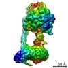
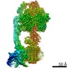
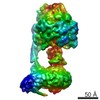
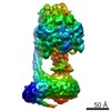
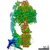
| Title | Structure of the mitochondrial ATP synthase from Saccharomyces cerevisiae in Brij-35 at 24 Angstroms resolution. | |||||||||
 Map data Map data | Map of the Saccharomyces cerevisiae mitochondrial ATP synthase in the detergent Brij-35 | |||||||||
 Sample Sample |
| |||||||||
 Keywords Keywords | ATP synthase / membrane protein / cryo-EM / detergent | |||||||||
| Biological species |  | |||||||||
| Method | single particle reconstruction / cryo EM / Resolution: 24.0 Å | |||||||||
 Authors Authors | Lau WCY / Baker LA / Rubinstein JL | |||||||||
 Citation Citation | Journal: J Mol Biol / Year: 2008 Title: Cryo-EM structure of the yeast ATP synthase. Authors: Wilson C Y Lau / Lindsay A Baker / John L Rubinstein /  Abstract: We have used electron cryomicroscopy of single particles to determine the structure of the ATP synthase from Saccharomyces cerevisiae. The resulting map at 24 A resolution can accommodate atomic ...We have used electron cryomicroscopy of single particles to determine the structure of the ATP synthase from Saccharomyces cerevisiae. The resulting map at 24 A resolution can accommodate atomic models of the F(1)-c(10) subcomplex, the peripheral stalk subcomplex, and the N-terminal domain of the oligomycin sensitivity conferral protein. The map is similar to an earlier electron cryomicroscopy structure of bovine mitochondrial ATP synthase but with important differences. It resolves the internal structure of the membrane region of the complex, especially the membrane embedded subunits b, c, and a. Comparison of the yeast ATP synthase map, which lacks density from the dimer-specific subunits e and g, with a map of the bovine enzyme that included e and g indicates where these subunits are located in the intact complex. This new map has allowed construction of a model of subunit arrangement in the F(O) motor of ATP synthase that dictates how dimerization of the complex via subunits e and g might occur. | |||||||||
| History |
|
- Structure visualization
| Movie |
 Movie viewer Movie viewer |
|---|---|
| Structure viewer | EM map: SurfViewMolmilJmol/JSmol |
| Supplemental images |

- Downloads & links
-EMDB archive
| Map data | emd_2011.map.gz | 949.1 KB | EMDB map data format | |
|---|---|---|---|---|
| Header (meta data) | emd-2011-v30.xmlemd-2011.xml | 9 KB 9 KB | Display Display | EMDB header |
| Images | 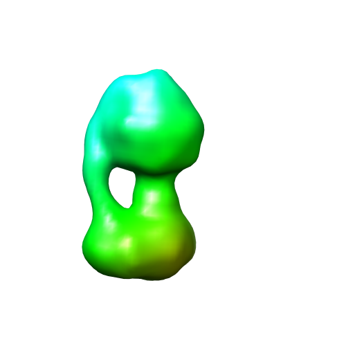 2011.png 2011.png | 53.7 KB | ||
| Archive directory |  http://ftp.pdbj.org/pub/emdb/structures/EMD-2011ftp://ftp.pdbj.org/pub/emdb/structures/EMD-2011 http://ftp.pdbj.org/pub/emdb/structures/EMD-2011ftp://ftp.pdbj.org/pub/emdb/structures/EMD-2011 | HTTPS FTP |
-Related structure data
| Similar structure data |
|---|
-Links
| EMDB pages | EMDB (EBI/PDBe) / EMDataResource |
|---|
-Map
| File | Download / File: emd_2011.map.gz / Format: CCP4 / Size: 1001 KB / Type: IMAGE STORED AS FLOATING POINT NUMBER (4 BYTES) | ||||||||||||||||||||||||||||||||||||||||||||||||||||||||||||||||||||
|---|---|---|---|---|---|---|---|---|---|---|---|---|---|---|---|---|---|---|---|---|---|---|---|---|---|---|---|---|---|---|---|---|---|---|---|---|---|---|---|---|---|---|---|---|---|---|---|---|---|---|---|---|---|---|---|---|---|---|---|---|---|---|---|---|---|---|---|---|---|
| Annotation | Map of the Saccharomyces cerevisiae mitochondrial ATP synthase in the detergent Brij-35 | ||||||||||||||||||||||||||||||||||||||||||||||||||||||||||||||||||||
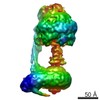
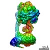
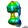
| Projections & slices | Image control
Images are generated by Spider. | ||||||||||||||||||||||||||||||||||||||||||||||||||||||||||||||||||||
| Voxel size | X=Y=Z: 5.6 Å | ||||||||||||||||||||||||||||||||||||||||||||||||||||||||||||||||||||
| Density |
| ||||||||||||||||||||||||||||||||||||||||||||||||||||||||||||||||||||
| Symmetry | Space group: 1 | ||||||||||||||||||||||||||||||||||||||||||||||||||||||||||||||||||||
| Details | EMDB XML:
CCP4 map header:
| ||||||||||||||||||||||||||||||||||||||||||||||||||||||||||||||||||||
 Z (Sec.)
 Y (Row.)
 X (Col.)


-Supplemental data
- Sample components
-Entire : Mitochondrial ATP synthase from Saccharomyces cerevisiae
| Entire | Name: Mitochondrial ATP synthase from Saccharomyces cerevisiae |
|---|---|
| Components |
|
-Supramolecule #1000: Mitochondrial ATP synthase from Saccharomyces cerevisiae
| Supramolecule | Name: Mitochondrial ATP synthase from Saccharomyces cerevisiae type: sample / ID: 1000 / Details: Sample contains Brij-35 to maintain solubility / Oligomeric state: One homo-oligomer / Number unique components: 14 |
|---|---|
| Molecular weight | Theoretical: 500 KDa |
-Macromolecule #1: ATP synthase
| Macromolecule | Name: ATP synthase / type: protein_or_peptide / ID: 1 / Name.synonym: ATP synthase / Oligomeric state: hetero-oligomer / Recombinant expression: Yes |
|---|---|
| Source (natural) | Organism: |
| Recombinant expression | Organism: |
-Experimental details
-Structure determination
| Method | cryo EM |
|---|---|
 Processing Processing | single particle reconstruction |
| Aggregation state | particle |
-Sample preparation
| Concentration | 3 mg/mL |
|---|---|
| Buffer | pH: 8 Details: 10 mM Tris-HCl, 10 mM NaCl, 2mM MgSO4, 0.05% (v/v) Brij35 |
| Grid | Details: 400 mesh Cu/Rh grid with homemade holey carbon |
| Vitrification | Cryogen name: ETHANE / Chamber humidity: 100 % / Instrument: FEI VITROBOT MARK III / Details: Vitrification instrument: Vitrobot Mark 3 / Method: Blot from both sides |
- Electron microscopy
| Microscope | FEI TECNAI F20 |
|---|---|
| Alignment procedure | Legacy - Astigmatism: Objective lens astigmatism corrected around 100,000x magnification |
| Image recording | Category: FILM / Film or detector model: KODAK SO-163 FILM / Digitization - Scanner: ZEISS SCAI / Digitization - Sampling interval: 7 µm / Number real images: 103 / Average electron dose: 12 e/Å2 / Od range: 1 / Bits/pixel: 8 |
| Electron beam | Acceleration voltage: 200 kV / Electron source:  FIELD EMISSION GUN FIELD EMISSION GUN |
| Electron optics | Calibrated magnification: 50000 / Illumination mode: FLOOD BEAM / Imaging mode: BRIGHT FIELD / Cs: 2.0 mm / Nominal defocus max: 6.0 µm / Nominal defocus min: 4.0 µm / Nominal magnification: 50000 |
| Sample stage | Specimen holder: Gatan 626 / Specimen holder model: SIDE ENTRY, EUCENTRIC |
| Experimental equipment |  Model: Tecnai F20 / Image courtesy: FEI Company |
-Image processing
| Details | particle selected manually |
|---|---|
| CTF correction | Details: Each particle |
| Final reconstruction | Applied symmetry - Point group: C1 (asymmetric) / Algorithm: OTHER / Resolution.type: BY AUTHOR / Resolution: 24.0 Å / Resolution method: FSC 0.143 CUT-OFF / Software - Name: Rotan, Spider, Frealign / Number images used: 6904 |
