Movie
Movie Controller


+ Open data

- Basic information
| Entry | Database: PDB / ID: 6tt7 | |||||||||
|---|---|---|---|---|---|---|---|---|---|---|
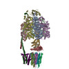
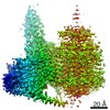
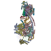
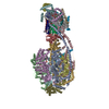
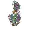
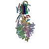
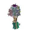
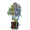
| Title | Ovine ATP synthase 1a state | |||||||||
 Components Components |
| |||||||||
 Keywords Keywords | MEMBRANE PROTEIN / ATP synthase / mitochondrial / respiratory chain / mammalian | |||||||||
| Function / homology |  Function and homology information Function and homology information: / : / negative regulation of cell adhesion involved in substrate-bound cell migration / : / angiostatin binding / Formation of ATP by chemiosmotic coupling / Cristae formation / mitochondrial proton-transporting ATP synthase complex assembly / cellular response to interleukin-7 / proton channel activity ...: / : / negative regulation of cell adhesion involved in substrate-bound cell migration / : / angiostatin binding / Formation of ATP by chemiosmotic coupling / Cristae formation / mitochondrial proton-transporting ATP synthase complex assembly / cellular response to interleukin-7 / proton channel activity / Mitochondrial protein degradation / proton transmembrane transporter activity / proton motive force-driven ATP synthesis / proton-transporting two-sector ATPase complex, proton-transporting domain / proton motive force-driven mitochondrial ATP synthesis / mitochondrial nucleoid / proton-transporting ATPase activity, rotational mechanism / MHC class I protein binding / positive regulation of blood vessel endothelial cell migration / H+-transporting two-sector ATPase / proton-transporting ATP synthase complex / proton-transporting ATP synthase activity, rotational mechanism / proton transmembrane transport / aerobic respiration / regulation of intracellular pH / lipid metabolic process / mitochondrial membrane / angiogenesis / mitochondrial inner membrane / lipid binding / cell surface / mitochondrion / ATP binding / plasma membrane Similarity search - Function | |||||||||
| Biological species |  | |||||||||
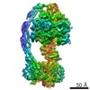
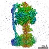
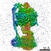
| Method | ELECTRON MICROSCOPY / single particle reconstruction / cryo EM / Resolution: 3.5 Å | |||||||||
 Authors Authors | Pinke, G. / Zhou, L. / Sazanov, L.A. | |||||||||
 Citation Citation | Journal: Nat Struct Mol Biol / Year: 2020 Title: Cryo-EM structure of the entire mammalian F-type ATP synthase. Authors: Gergely Pinke / Long Zhou / Leonid A Sazanov /  Abstract: The majority of adenosine triphosphate (ATP) powering cellular processes in eukaryotes is produced by the mitochondrial F1Fo ATP synthase. Here, we present the atomic models of the membrane Fo domain ...The majority of adenosine triphosphate (ATP) powering cellular processes in eukaryotes is produced by the mitochondrial F1Fo ATP synthase. Here, we present the atomic models of the membrane Fo domain and the entire mammalian (ovine) F1Fo, determined by cryo-electron microscopy. Subunits in the membrane domain are arranged in the 'proton translocation cluster' attached to the c-ring and a more distant 'hook apparatus' holding subunit e. Unexpectedly, this subunit is anchored to a lipid 'plug' capping the c-ring. We present a detailed proton translocation pathway in mammalian Fo and key inter-monomer contacts in F1Fo multimers. Cryo-EM maps of F1Fo exposed to calcium reveal a retracted subunit e and a disassembled c-ring, suggesting permeability transition pore opening. We propose a model for the permeability transition pore opening, whereby subunit e pulls the lipid plug out of the c-ring. Our structure will allow the design of drugs for many emerging applications in medicine. | |||||||||
| History |
|
- Structure visualization
| Movie |
Movie viewer |
|---|---|
| Structure viewer | Molecule: MolmilJmol/JSmol |
- Downloads & links
-Download
| PDBx/mmCIF format | 6tt7.cif.gz | 1 MB | Display | PDBx/mmCIF format |
|---|---|---|---|---|
| PDB format | pdb6tt7.ent.gz | 825 KB | Display | PDB format |
| PDBx/mmJSON format | 6tt7.json.gz | Tree view | PDBx/mmJSON format | |
| Others |  Other downloads Other downloads |
-Validation report
| Arichive directory | https://data.pdbj.org/pub/pdb/validation_reports/tt/6tt7ftp://data.pdbj.org/pub/pdb/validation_reports/tt/6tt7 | HTTPS FTP |
|---|
-Related structure data
| Related structure data | 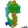 10573MC  6za9C M: map data used to model this data C: citing same article ( |
|---|---|
| Similar structure data |
-Links
 PDBj
PDBj


- Assembly
| Deposited unit | 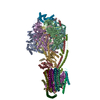
|
|---|---|
| 1 |
|
-Components
-ATP synthase ... , 14 types, 25 molecules BCAEDFGHIJKM12345678NPQRS
| #1: Protein | Mass: 59779.492 Da / Num. of mol.: 3 / Source method: isolated from a natural source / Source: (natural) #2: Protein | Mass: 56265.090 Da / Num. of mol.: 3 / Source method: isolated from a natural source / Source: (natural) References: UniProt: W5PEP7, H+-transporting two-sector ATPase #3: Protein | | Mass: 33133.062 Da / Num. of mol.: 1 / Source method: isolated from a natural source / Details: XP_004014229.1 / Source: (natural) #4: Protein | | Mass: 17580.924 Da / Num. of mol.: 1 / Source method: isolated from a natural source / Source: (natural) #5: Protein | | Mass: 5779.863 Da / Num. of mol.: 1 / Source method: isolated from a natural source / Details: XP_027832792.1 / Source: (natural) #6: Protein | | Mass: 23435.695 Da / Num. of mol.: 1 / Source method: isolated from a natural source / Source: (natural) #7: Protein | | Mass: 28844.570 Da / Num. of mol.: 1 / Source method: isolated from a natural source / Details: XP_004002367.2 / Source: (natural) #9: Protein | | Mass: 18747.508 Da / Num. of mol.: 1 / Source method: isolated from a natural source / Source: (natural) #10: Protein | Mass: 14201.577 Da / Num. of mol.: 8 / Source method: isolated from a natural source / Source: (natural) #11: Protein | | Mass: 24811.850 Da / Num. of mol.: 1 / Source method: isolated from a natural source / Source: (natural) #13: Protein | | Mass: 6846.093 Da / Num. of mol.: 1 / Source method: isolated from a natural source / Source: (natural) #14: Protein | | Mass: 7918.443 Da / Num. of mol.: 1 / Source method: isolated from a natural source / Source: (natural) #15: Protein | | Mass: 10315.207 Da / Num. of mol.: 1 / Source method: isolated from a natural source / Details: XP_004016938.3 / Source: (natural) #16: Protein | | Mass: 11428.407 Da / Num. of mol.: 1 / Source method: isolated from a natural source / Details: XP_011950914.1 / Source: (natural) |
|---|
-Protein , 3 types, 3 molecules LOT
| #8: Protein | Mass: 12490.296 Da / Num. of mol.: 1 / Source method: isolated from a natural source / Source: (natural) |
|---|---|
| #12: Protein | Mass: 6443.579 Da / Num. of mol.: 1 / Source method: isolated from a natural source / Source: (natural) |
| #17: Protein | Mass: 8336.688 Da / Num. of mol.: 1 / Source method: isolated from a natural source / Details: XP_027827162.1 / Source: (natural) |
-Non-polymers , 6 types, 19 molecules 


| #18: Chemical |  Mass: 24.305 Da / Num. of mol.: 3 / Source method: obtained synthetically / Formula: Mg Mass: 24.305 Da / Num. of mol.: 3 / Source method: obtained synthetically / Formula: Mg#19: Chemical | ChemComp-ADP /  Mass: 427.201 Da / Num. of mol.: 5 / Source method: obtained synthetically / Formula: C10H15N5O10P2 / Comment: ADP, energy-carrying molecule*YM Mass: 427.201 Da / Num. of mol.: 5 / Source method: obtained synthetically / Formula: C10H15N5O10P2 / Comment: ADP, energy-carrying molecule*YM#20: Chemical | ChemComp-LHG /  Mass: 722.970 Da / Num. of mol.: 6 / Source method: obtained synthetically / Formula: C38H75O10P / Comment: phospholipid*YM Mass: 722.970 Da / Num. of mol.: 6 / Source method: obtained synthetically / Formula: C38H75O10P / Comment: phospholipid*YM#21: Chemical | ChemComp-P5S / |  Mass: 792.075 Da / Num. of mol.: 1 / Source method: obtained synthetically / Formula: C42H82NO10P Mass: 792.075 Da / Num. of mol.: 1 / Source method: obtained synthetically / Formula: C42H82NO10P#22: Chemical |  Mass: 1464.043 Da / Num. of mol.: 3 / Source method: obtained synthetically / Formula: C81H156O17P2 / Comment: phospholipid*YM Mass: 1464.043 Da / Num. of mol.: 3 / Source method: obtained synthetically / Formula: C81H156O17P2 / Comment: phospholipid*YM#23: Chemical | ChemComp-S12 / |  Type: L-peptide linking / Mass: 523.597 Da / Num. of mol.: 1 / Source method: obtained synthetically / Formula: C24H46NO9P Type: L-peptide linking / Mass: 523.597 Da / Num. of mol.: 1 / Source method: obtained synthetically / Formula: C24H46NO9P |
|---|
-Details
| Has ligand of interest | N |
|---|
-Experimental details
-Experiment
| Experiment | Method: ELECTRON MICROSCOPY |
|---|---|
| EM experiment | Aggregation state: PARTICLE / 3D reconstruction method: single particle reconstruction |
- Sample preparation
| Component | Name: Purified ATP synthase from sheep heart mitochondria / Type: COMPLEX / Entity ID: #1-#17 / Source: NATURAL |
|---|---|
| Molecular weight | Experimental value: NO |
| Source (natural) | Organism: |
| Buffer solution | pH: 7.4 Details: 20 mM HEPES pH7.4, 35 mM NaCl, 2 mM EDTA, 1% sucrose, 1 mM DTT, 0.1% LMNG, 0.2% CHAPS |
| Specimen | Conc.: 2.8 mg/ml / Embedding applied: NO / Shadowing applied: NO / Staining applied: NO / Vitrification applied: YES |
| Specimen support | Grid material: COPPER / Grid mesh size: 400 divisions/in. / Grid type: Quantifoil R0.6/1 |
| Vitrification | Instrument: FEI VITROBOT MARK IV / Cryogen name: ETHANE-PROPANE / Humidity: 100 % / Chamber temperature: 277 K |
- Electron microscopy imaging
| Experimental equipment |  Model: Titan Krios / Image courtesy: FEI Company |
|---|---|
| Microscopy | Model: FEI TITAN KRIOS |
| Electron gun | Electron source:  FIELD EMISSION GUN / Accelerating voltage: 300 kV / Illumination mode: FLOOD BEAM FIELD EMISSION GUN / Accelerating voltage: 300 kV / Illumination mode: FLOOD BEAM |
| Electron lens | Mode: BRIGHT FIELD / Calibrated magnification: 131951 X / Calibrated defocus min: 1000 nm / Calibrated defocus max: 2400 nm / Cs: 2.7 mm / Alignment procedure: COMA FREE |
| Specimen holder | Cryogen: NITROGEN / Specimen holder model: FEI TITAN KRIOS AUTOGRID HOLDER |
| Image recording | Average exposure time: 1 sec. / Electron dose: 106 e/Å2 / Detector mode: INTEGRATING / Film or detector model: FEI FALCON III (4k x 4k) / Num. of grids imaged: 1 / Num. of real images: 3053 / Details: 40 frames per movie |
| Image scans | Sampling size: 14 µm / Width: 4096 / Height: 4096 |
- Processing
| Software | Name: PHENIX / Version: 1.12_2829: / Classification: refinement | ||||||||||||||||||||||||||||||||||||||||||||||||||||||||||||
|---|---|---|---|---|---|---|---|---|---|---|---|---|---|---|---|---|---|---|---|---|---|---|---|---|---|---|---|---|---|---|---|---|---|---|---|---|---|---|---|---|---|---|---|---|---|---|---|---|---|---|---|---|---|---|---|---|---|---|---|---|---|
| EM software |
| ||||||||||||||||||||||||||||||||||||||||||||||||||||||||||||
| CTF correction | Type: PHASE FLIPPING AND AMPLITUDE CORRECTION | ||||||||||||||||||||||||||||||||||||||||||||||||||||||||||||
| Particle selection | Num. of particles selected: 809000 | ||||||||||||||||||||||||||||||||||||||||||||||||||||||||||||
| Symmetry | Point symmetry: C1 (asymmetric) | ||||||||||||||||||||||||||||||||||||||||||||||||||||||||||||
| 3D reconstruction | Resolution: 3.5 Å / Resolution method: FSC 0.143 CUT-OFF / Num. of particles: 35000 / Symmetry type: POINT | ||||||||||||||||||||||||||||||||||||||||||||||||||||||||||||
| Atomic model building |
| ||||||||||||||||||||||||||||||||||||||||||||||||||||||||||||
| Atomic model building |
| ||||||||||||||||||||||||||||||||||||||||||||||||||||||||||||
| Refine LS restraints |
|