 Movie
Movie Controller

[English] 日本語
 Yorodumi
- PDB-3j09: High resolution helical reconstruction of the bacterial p-type AT... -
+ Open data

- Basic information
| Entry | Database: PDB / ID: 3j09 | ||||||
|---|---|---|---|---|---|---|---|
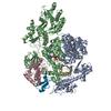
| Title | High resolution helical reconstruction of the bacterial p-type ATPase copper transporter CopA | ||||||
 Components Components | copper-exporting P-type ATPase A | ||||||
 Keywords Keywords | HYDROLASE / METAL TRANSPORT / p-type ATPase / copper transporter / CopA / adenosine triphosphatases / archaeal proteins / cation transport proteins / cryoelectron microscopy | ||||||
| Function / homology |  Function and homology information Function and homology informationP-type divalent copper transporter activity / P-type Cu+ transporter / P-type monovalent copper transporter activity / copper ion homeostasis / copper ion binding / ATP hydrolysis activity / ATP binding / identical protein binding / plasma membrane Similarity search - Function | ||||||
| Biological species |   Archaeoglobus fulgidus (archaea) Archaeoglobus fulgidus (archaea) | ||||||
| Method | ELECTRON MICROSCOPY / helical reconstruction / cryo EM / Resolution: 10 Å | ||||||
 Authors Authors | Wu, C. / Allen, G.S. / Cardozo, T. / Stokes, D.L. | ||||||
 Citation Citation | Journal: Structure / Year: 2011 Title: The architecture of CopA from Archeaoglobus fulgidus studied by cryo-electron microscopy and computational docking. Authors: Gregory S Allen / Chen-Chou Wu / Tim Cardozo / David L Stokes /  Abstract: CopA uses ATP to pump Cu(+) across cell membranes. X-ray crystallography has defined atomic structures of several related P-type ATPases. We have determined a structure of CopA at 10 Å resolution ...CopA uses ATP to pump Cu(+) across cell membranes. X-ray crystallography has defined atomic structures of several related P-type ATPases. We have determined a structure of CopA at 10 Å resolution by cryo-electron microscopy of a new crystal form and used computational molecular docking to study the interactions between the N-terminal metal-binding domain (NMBD) and other elements of the molecule. We found that the shorter-chain lipids used to produce these crystals are associated with movements of the cytoplasmic domains, with a novel dimer interface and with disordering of the NMBD, thus offering evidence for the transience of its interaction with the other cytoplasmic domains. Docking identified a binding site that matched the location of the NMBD in our previous structure by cryo-electron microscopy, allowing a more detailed view of its binding configuration and further support for its role in autoinhibition. | ||||||
| History |
|
- Structure visualization
| Movie |
Movie viewer |
|---|---|
| Structure viewer | Molecule: MolmilJmol/JSmol |
- Downloads & links
-Download
| PDBx/mmCIF format | 3j09.cif.gz | 246 KB | Display | PDBx/mmCIF format |
|---|---|---|---|---|
| PDB format | pdb3j09.ent.gz | 188.5 KB | Display | PDB format |
| PDBx/mmJSON format | 3j09.json.gz | Tree view | PDBx/mmJSON format | |
| Others |  Other downloads Other downloads |
-Validation report
| Arichive directory | https://data.pdbj.org/pub/pdb/validation_reports/j0/3j09ftp://data.pdbj.org/pub/pdb/validation_reports/j0/3j09 | HTTPS FTP |
|---|
-Related structure data
| Related structure data |  5004M 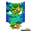 5271C  3j08C C: citing same article ( M: map data used to model this data |
|---|---|
| Similar structure data |


-Links
 PDBj
PDBj


- Assembly
| Deposited unit | 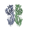
|
|---|---|
| 1 |
|
-Components
| #1: Protein | Mass: 77815.648 Da / Num. of mol.: 2 / Fragment: deltaC-CopA (UNP residues 15-737) Source method: isolated from a genetically manipulated source Source: (gene. exp.) Archaeoglobus fulgidus (archaea) / Gene: copA, pacS, AF_0473 / Production host:  References: UniProt: O29777, Hydrolases; Acting on acid anhydrides; Acting on acid anhydrides to catalyse transmembrane movement of substances |
|---|
-Experimental details
-Experiment
| Experiment | Method: ELECTRON MICROSCOPY |
|---|---|
| EM experiment | Aggregation state: HELICAL ARRAY / 3D reconstruction method: helical reconstruction |
- Sample preparation
| Component | Name: deltaC-CopA in DOPC lipids / Type: COMPLEX Details: DeltaC-CopA tubular crystals were grown with DOPC at a protein concentration of 1 mg/mL and at a lipid-to-protein weight ratio of 0.4. Dialysis was carried out for 5 days in 50 uL dialysis ...Details: DeltaC-CopA tubular crystals were grown with DOPC at a protein concentration of 1 mg/mL and at a lipid-to-protein weight ratio of 0.4. Dialysis was carried out for 5 days in 50 uL dialysis buttons at 303K against 500 mL of 50 mM MES, pH 6.1, 25 mM Na2SO4, 25 mM K2SO4, 200 uM BCDS, 10 mM MgSO4, and 2 mM beta-mercaptoethanol. Stock solutions of lipid were made in dodecyl octaethylene glycol ether (C12E8) at 1 mg lipid per 2 mg detergent. |
|---|---|
| Molecular weight | Value: 0.077 MDa / Experimental value: NO |
| Buffer solution | pH: 6.1 Details: 50 mM MES, pH 6.1, 25 mM Na2SO4, 25 mM K2SO4, 200 uM BCDS, 10 mM MgSO4, 2 mM beta-mercaptoethanol |
| Specimen | Conc.: 1 mg/ml / Embedding applied: NO / Shadowing applied: NO / Staining applied: NO / Vitrification applied: YES Details: 50 mM MES, pH 6.1, 25 mM Na2SO4, 25 mM K2SO4, 200 uM BCDS, 10 mM MgSO4, 2 mM beta-mercaptoethanol |
| Vitrification | Instrument: HOMEMADE PLUNGER / Cryogen name: ETHANE / Temp: 77 K / Method: blot for 5 seconds before plunging |
- Electron microscopy imaging
| Microscopy | Model: FEI/PHILIPS CM200FEG / Date: Jan 1, 2009 |
|---|---|
| Electron gun | Electron source:  FIELD EMISSION GUN / Accelerating voltage: 200 kV / Illumination mode: SPOT SCAN FIELD EMISSION GUN / Accelerating voltage: 200 kV / Illumination mode: SPOT SCAN |
| Electron lens | Mode: BRIGHT FIELD / Nominal magnification: 50000 X / Nominal defocus max: 2500 nm / Nominal defocus min: 1500 nm / Cs: 2 mm / Camera length: 0 mm |
| Specimen holder | Specimen holder model: GATAN LIQUID NITROGEN / Specimen holder type: CT3500 / Temperature: 100 K / Tilt angle max: 0 ° / Tilt angle min: 0 ° |
| Image recording | Electron dose: 10 e/Å2 / Film or detector model: KODAK SO-163 FILM |
| Radiation | Protocol: SINGLE WAVELENGTH / Monochromatic (M) / Laue (L): M / Scattering type: x-ray |
| Radiation wavelength | Relative weight: 1 |
- Processing
| EM software | Name: EMIP / Category: 3D reconstruction | ||||||||||||
|---|---|---|---|---|---|---|---|---|---|---|---|---|---|
| CTF correction | Details: each tube-crystal | ||||||||||||
| 3D reconstruction | Method: Fourier-Bessel / Resolution: 10 Å / Resolution method: OTHER / Symmetry type: HELICAL | ||||||||||||
| Refinement step | Cycle: LAST
|
