 Movie
Movie Controller

[English] 日本語
 Yorodumi
- EMDB-5271: High resolution helical reconstruction of the bacterial p-type AT... -
+ Open data

- Basic information
| Entry | Database: EMDB / ID: EMD-5271 | |||||||||
|---|---|---|---|---|---|---|---|---|---|---|
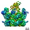
| Title | High resolution helical reconstruction of the bacterial p-type ATPase copper transporter CopA. | |||||||||
 Map data Map data | This is a cubic section encompassing the unit cell of a Fourier-Bessel reconstructed map of tubular vesicles - crystals of deltaC-CopA from Archaeoglobus fulgidus. | |||||||||
 Sample Sample |
| |||||||||
 Keywords Keywords | p-type ATPase / copper transporter / CopA / Adenosine Triphosphatases / Archaeal Proteins / Archaeoglobus fulgidus / Cation Transport Proteins / Cryoelectron Microscopy | |||||||||
| Function / homology |  Function and homology information Function and homology informationP-type divalent copper transporter activity / P-type Cu+ transporter / P-type monovalent copper transporter activity / copper ion homeostasis / copper ion transport / copper ion binding / ATP hydrolysis activity / ATP binding / identical protein binding / plasma membrane Similarity search - Function | |||||||||
| Biological species |   Archaeoglobus fulgidus (archaea) Archaeoglobus fulgidus (archaea) | |||||||||
| Method | helical reconstruction / cryo EM / Resolution: 10.0 Å | |||||||||
 Authors Authors | Wu C / Allen GS / Cardozo T / Stokes DL | |||||||||
 Citation Citation | Journal: Structure / Year: 2011 Title: The architecture of CopA from Archeaoglobus fulgidus studied by cryo-electron microscopy and computational docking. Authors: Gregory S Allen / Chen-Chou Wu / Tim Cardozo / David L Stokes /  Abstract: CopA uses ATP to pump Cu(+) across cell membranes. X-ray crystallography has defined atomic structures of several related P-type ATPases. We have determined a structure of CopA at 10 Å resolution ...CopA uses ATP to pump Cu(+) across cell membranes. X-ray crystallography has defined atomic structures of several related P-type ATPases. We have determined a structure of CopA at 10 Å resolution by cryo-electron microscopy of a new crystal form and used computational molecular docking to study the interactions between the N-terminal metal-binding domain (NMBD) and other elements of the molecule. We found that the shorter-chain lipids used to produce these crystals are associated with movements of the cytoplasmic domains, with a novel dimer interface and with disordering of the NMBD, thus offering evidence for the transience of its interaction with the other cytoplasmic domains. Docking identified a binding site that matched the location of the NMBD in our previous structure by cryo-electron microscopy, allowing a more detailed view of its binding configuration and further support for its role in autoinhibition. | |||||||||
| History |
|
- Structure visualization
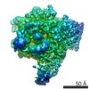
| Movie |
Movie viewer |
|---|---|
| Structure viewer | EM map: SurfViewMolmilJmol/JSmol |
| Supplemental images |

- Downloads & links
-EMDB archive
| Map data | emd_5271.map.gz | 677.1 KB | EMDB map data format | |
|---|---|---|---|---|
| Header (meta data) | emd-5271-v30.xmlemd-5271.xml | 10.2 KB 10.2 KB | Display Display | EMDB header |
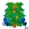
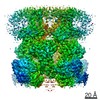
| Images | 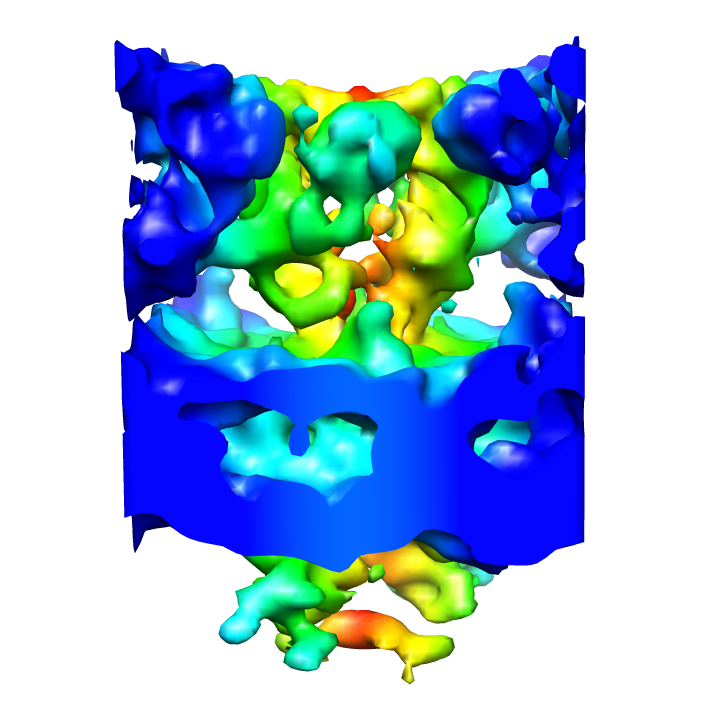 emd_5271_1.png emd_5271_1.png | 220.1 KB | ||
| Archive directory |  http://ftp.pdbj.org/pub/emdb/structures/EMD-5271ftp://ftp.pdbj.org/pub/emdb/structures/EMD-5271 http://ftp.pdbj.org/pub/emdb/structures/EMD-5271ftp://ftp.pdbj.org/pub/emdb/structures/EMD-5271 | HTTPS FTP |
-Related structure data
| Related structure data |  3j08MC 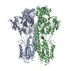 3j09C M: atomic model generated by this map C: citing same article ( |
|---|---|
| Similar structure data |


-Links
| EMDB pages | EMDB (EBI/PDBe) / EMDataResource |
|---|---|
| Related items in Molecule of the Month |


-Map
| File | Download / File: emd_5271.map.gz / Format: CCP4 / Size: 789.1 KB / Type: IMAGE STORED AS FLOATING POINT NUMBER (4 BYTES) | ||||||||||||||||||||||||||||||||||||||||||||||||||||||||||||||||||||
|---|---|---|---|---|---|---|---|---|---|---|---|---|---|---|---|---|---|---|---|---|---|---|---|---|---|---|---|---|---|---|---|---|---|---|---|---|---|---|---|---|---|---|---|---|---|---|---|---|---|---|---|---|---|---|---|---|---|---|---|---|---|---|---|---|---|---|---|---|---|
| Annotation | This is a cubic section encompassing the unit cell of a Fourier-Bessel reconstructed map of tubular vesicles - crystals of deltaC-CopA from Archaeoglobus fulgidus. | ||||||||||||||||||||||||||||||||||||||||||||||||||||||||||||||||||||
| Projections & slices | Image control
Images are generated by Spider. generated in cubic-lattice coordinate | ||||||||||||||||||||||||||||||||||||||||||||||||||||||||||||||||||||
| Voxel size | X=Y=Z: 2 Å | ||||||||||||||||||||||||||||||||||||||||||||||||||||||||||||||||||||
| Density |
| ||||||||||||||||||||||||||||||||||||||||||||||||||||||||||||||||||||
| Symmetry | Space group: 1 | ||||||||||||||||||||||||||||||||||||||||||||||||||||||||||||||||||||
| Details | EMDB XML:
CCP4 map header:
| ||||||||||||||||||||||||||||||||||||||||||||||||||||||||||||||||||||
 Z (Sec.)
 Y (Row.)
 X (Col.)


-Supplemental data
- Sample components
-Entire : deltaC-CopA in DMPC-DOPE lipids
| Entire | Name: deltaC-CopA in DMPC-DOPE lipids |
|---|---|
| Components |
|
-Supramolecule #1000: deltaC-CopA in DMPC-DOPE lipids
| Supramolecule | Name: deltaC-CopA in DMPC-DOPE lipids / type: sample / ID: 1000 Details: deltaC-CopA tubular crystals were grown with a 4-to-1 mixture of DMPC-DOPE at a protein concentration of 1 mg per mL and at a lipid-to-protein weight ratio of 0.4. Dialysis was carried out ...Details: deltaC-CopA tubular crystals were grown with a 4-to-1 mixture of DMPC-DOPE at a protein concentration of 1 mg per mL and at a lipid-to-protein weight ratio of 0.4. Dialysis was carried out for 5 days in 50 ul dialysis buttons at 30 degrees C against 500 mL of 50 mM MES, pH 6.1, 25 mM Na2SO4, 25 mM K2SO4, 200 uM BCDS, 10 mM MgSO4, and 2 mM beta-mercaptoethanol. Stock solutions of lipid were made in dodecyl octaethylene glycol ether (C12E8) at 1 mg lipid per 2 mg detergent. Number unique components: 1 |
|---|---|
| Molecular weight | Theoretical: 77 KDa |
-Macromolecule #1: membrane protein
| Macromolecule | Name: membrane protein / type: protein_or_peptide / ID: 1 / Name.synonym: membrane protein / Number of copies: 2 / Oligomeric state: dimer / Recombinant expression: Yes |
|---|---|
| Source (natural) | Organism: Archaeoglobus fulgidus (archaea) / Cell: E. coli / Location in cell: plasma membrane |
| Molecular weight | Theoretical: 77 KDa |
| Recombinant expression | Organism: Escherichia coli strain LMG 1940 / Recombinant plasmid: pBAD |
| Sequence | GO: copper ion transport / InterPro: INTERPRO: IPR006403 |
-Experimental details
-Structure determination
| Method | cryo EM |
|---|---|
 Processing Processing | helical reconstruction |
| Aggregation state | helical array |
-Sample preparation
| Concentration | 1 mg/mL |
|---|---|
| Buffer | pH: 6.1 Details: 50 mM MES, pH 6.1, 25 mM Na2SO4, 25 mM K2SO4, 200 uM BCDS, 10 mM MgSO4, and 2 mM beta-mercaptoethanol. |
| Grid | Details: holey carbon grid |
| Vitrification | Cryogen name: ETHANE / Chamber temperature: 77 K / Instrument: HOMEMADE PLUNGER / Details: Vitrification instrument: plunger / Method: blot for 5 seconds before plunging |
| Details | dialysis buttons |
- Electron microscopy
| Microscope | FEI/PHILIPS CM200FEG |
|---|---|
| Temperature | Average: 100 K |
| Image recording | Category: FILM / Film or detector model: KODAK SO-163 FILM / Digitization - Scanner: ZEISS SCAI / Digitization - Sampling interval: 14 µm / Number real images: 12 |
| Electron beam | Acceleration voltage: 200 kV / Electron source:  FIELD EMISSION GUN FIELD EMISSION GUN |
| Electron optics | Illumination mode: SPOT SCAN / Imaging mode: BRIGHT FIELD / Cs: 2 mm / Nominal defocus max: 2.5 µm / Nominal defocus min: 1.5 µm / Nominal magnification: 50000 |
| Sample stage | Specimen holder: CT3500 / Specimen holder model: GATAN LIQUID NITROGEN |
-Image processing
| Final reconstruction | Algorithm: OTHER / Resolution.type: BY AUTHOR / Resolution: 10.0 Å / Resolution method: OTHER / Software - Name: EMIP |
|---|---|
| CTF correction | Details: each tube-crystal |
