Type: RIGAKU RAXIS IV / Detector: IMAGE PLATE / Date: Jun 26, 2010
Radiation
Protocol: SINGLE WAVELENGTH / Monochromatic (M) / Laue (L): M / Scattering type: x-ray
Radiation wavelength
Wavelength: 1.52 Å / Relative weight: 1
Reflection
Redundancy: 6.5 % / Av σ(I) over netI: 4 / Number: 60901 / Rsym value: 0.185 / D res high: 1.975 Å / D res low: 28.641 Å / Num. obs: 9376 / % possible obs: 98.8
Diffraction reflection shell
Highest resolution (Å)
Lowest resolution (Å)
% possible obs (%)
ID
Rmerge(I) obs
Rsym value
Redundancy
6.25
28.64
97.9
1
0.065
0.065
5.8
4.42
6.25
100
1
0.088
0.088
6.5
3.61
4.42
99.9
1
0.088
0.088
6.6
3.12
3.61
99.6
1
0.123
0.123
6.8
2.79
3.12
99.7
1
0.202
0.202
6.7
2.55
2.79
99.4
1
0.282
0.282
6.8
2.36
2.55
98.9
1
0.359
0.359
6.8
2.21
2.36
98.8
1
0.441
0.441
6.9
2.08
2.21
98.4
1
0.527
0.527
6.8
1.98
2.08
96.4
1
0.745
0.745
5.2
Reflection
Resolution: 1.975→28.641 Å / Num. all: 9490 / Num. obs: 9376 / % possible obs: 98.8 % / Redundancy: 6.5 % / Biso Wilson estimate: 20 Å2 / Rmerge(I) obs: 0.185 / Rsym value: 0.185 / Net I/σ(I): 7.5
Reflection shell
Diffraction-ID: 1
Resolution (Å)
Redundancy (%)
Rmerge(I) obs
Mean I/σ(I) obs
Num. measured all
Num. unique all
Rsym value
% possible all
1.975-2.08
5.2
0.745
1
6720
1302
0.745
96.4
2.08-2.21
6.8
0.527
1.5
8559
1257
0.527
98.4
2.21-2.36
6.9
0.441
1.8
8175
1193
0.441
98.8
2.36-2.55
6.8
0.359
2.2
7594
1122
0.359
98.9
2.55-2.79
6.8
0.282
2.7
7078
1039
0.282
99.4
2.79-3.12
6.7
0.202
3.8
6513
967
0.202
99.7
3.12-3.61
6.8
0.123
6
5653
837
0.123
99.6
3.61-4.42
6.6
0.088
8.2
4823
729
0.088
99.9
4.42-6.25
6.5
0.088
7.9
3817
590
0.088
100
6.25-28.641
5.8
0.065
10.3
1969
340
0.065
97.9
-
Phasing
Phasing
Method: molecular replacement
Phasing MR
Rfactor: 38.14 / Model details: Phaser MODE: MR_AUTO
Resolution: 1.975→28.64 Å / Cor.coef. Fo:Fc: 0.95 / Cor.coef. Fo:Fc free: 0.922 / WRfactor Rfree: 0.201 / WRfactor Rwork: 0.1561 / Occupancy max: 1 / Occupancy min: 1 / FOM work R set: 0.8696 / SU B: 7.758 / SU ML: 0.115 / SU R Cruickshank DPI: 0.1873 / SU Rfree: 0.1656 / Cross valid method: THROUGHOUT / σ(F): 0 / ESU R Free: 0.166 / Stereochemistry target values: MAXIMUM LIKELIHOOD Details: HYDROGENS HAVE BEEN USED IF PRESENT IN THE INPUT. U VALUES WITH TLS ADDED.
Rfactor
Num. reflection
% reflection
Selection details
Rfree
0.2265
473
5.1 %
RANDOM
Rwork
0.1767
-
-
-
all
0.1791
9492
-
-
obs
0.1791
9347
98.47 %
-
Solvent computation
Ion probe radii: 0.8 Å / Shrinkage radii: 0.8 Å / VDW probe radii: 1.2 Å / Solvent model: MASK
In the structure databanks used in Yorodumi, some data are registered as the other names, "COVID-19 virus" and "2019-nCoV". Here are the details of the virus and the list of structure data.
Jan 31, 2019. EMDB accession codes are about to change! (news from PDBe EMDB page)
EMDB accession codes are about to change! (news from PDBe EMDB page)
The allocation of 4 digits for EMDB accession codes will soon come to an end. Whilst these codes will remain in use, new EMDB accession codes will include an additional digit and will expand incrementally as the available range of codes is exhausted. The current 4-digit format prefixed with “EMD-” (i.e. EMD-XXXX) will advance to a 5-digit format (i.e. EMD-XXXXX), and so on. It is currently estimated that the 4-digit codes will be depleted around Spring 2019, at which point the 5-digit format will come into force.
The EM Navigator/Yorodumi systems omit the EMD- prefix.
Related info.:Q: What is EMD? / ID/Accession-code notation in Yorodumi/EM Navigator
Yorodumi is a browser for structure data from EMDB, PDB, SASBDB, etc.
This page is also the successor to EM Navigator detail page, and also detail information page/front-end page for Omokage search.
The word "yorodu" (or yorozu) is an old Japanese word meaning "ten thousand". "mi" (miru) is to see.
Related info.:EMDB / PDB / SASBDB / Comparison of 3 databanks / Yorodumi Search / Aug 31, 2016. New EM Navigator & Yorodumi / Yorodumi Papers / Jmol/JSmol / Function and homology information / Changes in new EM Navigator and Yorodumi
 Movie
Movie Controller

 Yorodumi
 Open data
 Basic information
 Components
 Keywords
 Function and homology information
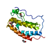
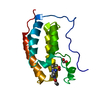
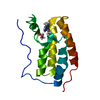
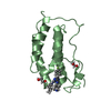
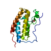
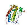
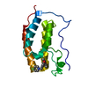
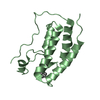
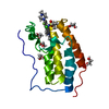
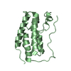
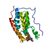
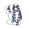
 Homo sapiens (human)
 X-RAY DIFFRACTION /
  Authors
 Citation
 Structure visualization
 Downloads & links
 Other downloads
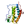


 PDBj
PDBj Assembly
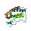

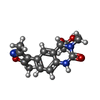
 Mass: 317.340 Da / Num. of mol.: 1 / Source method: obtained synthetically / Formula: C16H19N3O4
 Mass: 18.015 Da / Num. of mol.: 86 / Source method: isolated from a natural source / Formula: H2O
 Sample preparation
 Processing