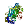
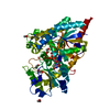
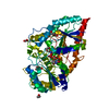
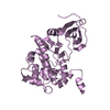
- PDB-3spw: Structure of Osh4p/Kes1p in complex with phosphatidylinositol 4-p... -
+
Open data
ID or keywords:
Loading...
-
Basic information
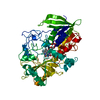
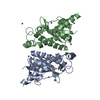
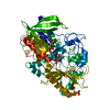
Entry
Database: PDB / ID: 3spw
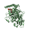
Title
Structure of Osh4p/Kes1p in complex with phosphatidylinositol 4-phosphate
Components
Protein KES1
Keywords
PROTEIN BINDING / Lipid binding protein
Function / homology
Function and homology information
Acyl chain remodelling of PS / Synthesis of bile acids and bile salts / ER to Golgi ceramide transport / sterol transfer activity / post-Golgi vesicle-mediated transport / sterol transport / sphingolipid metabolic process / maintenance of cell polarity / piecemeal microautophagy of the nucleus / oxysterol binding ...Acyl chain remodelling of PS / Synthesis of bile acids and bile salts / ER to Golgi ceramide transport / sterol transfer activity / post-Golgi vesicle-mediated transport / sterol transport / sphingolipid metabolic process / maintenance of cell polarity / piecemeal microautophagy of the nucleus / oxysterol binding / phosphatidic acid binding / phosphatidylinositol-4-phosphate binding / exocytosis / phosphatidylinositol-4,5-bisphosphate binding / endocytosis / Golgi membrane / lipid binding / membrane / cytosol / cytoplasm Similarity search - Function
Helix Hairpins - #1150 / Oxysterol-binding protein / Oxysterol-binding protein, conserved site / Oxysterol-binding protein superfamily / Oxysterol-binding protein / Oxysterol-binding protein family signature. / Helix Hairpins / Helix non-globular / Special Similarity search - Domain/homology
In the structure databanks used in Yorodumi, some data are registered as the other names, "COVID-19 virus" and "2019-nCoV". Here are the details of the virus and the list of structure data.
Jan 31, 2019. EMDB accession codes are about to change! (news from PDBe EMDB page)
EMDB accession codes are about to change! (news from PDBe EMDB page)
The allocation of 4 digits for EMDB accession codes will soon come to an end. Whilst these codes will remain in use, new EMDB accession codes will include an additional digit and will expand incrementally as the available range of codes is exhausted. The current 4-digit format prefixed with “EMD-” (i.e. EMD-XXXX) will advance to a 5-digit format (i.e. EMD-XXXXX), and so on. It is currently estimated that the 4-digit codes will be depleted around Spring 2019, at which point the 5-digit format will come into force.
The EM Navigator/Yorodumi systems omit the EMD- prefix.
Related info.:Q: What is EMD? / ID/Accession-code notation in Yorodumi/EM Navigator
Yorodumi is a browser for structure data from EMDB, PDB, SASBDB, etc.
This page is also the successor to EM Navigator detail page, and also detail information page/front-end page for Omokage search.
The word "yorodu" (or yorozu) is an old Japanese word meaning "ten thousand". "mi" (miru) is to see.
Related info.:EMDB / PDB / SASBDB / Comparison of 3 databanks / Yorodumi Search / Aug 31, 2016. New EM Navigator & Yorodumi / Yorodumi Papers / Jmol/JSmol / Function and homology information / Changes in new EM Navigator and Yorodumi
 Movie
Movie Controller

 Yorodumi
 Open data
 Basic information
 Components
 Keywords
 Function and homology information
 X-RAY DIFFRACTION /
  Authors
 Citation
 Structure visualization
 Downloads & links
 Other downloads


 PDBj
PDBj Assembly
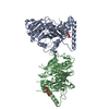


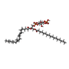
 Mass: 953.081 Da / Num. of mol.: 2 / Source method: obtained synthetically / Formula: C46H82O16P2
 Mass: 18.015 Da / Num. of mol.: 241 / Source method: isolated from a natural source / Formula: H2O
 Sample preparation
 / Beamline: ID14-1 / Wavelength: 0.9334 Å
 Processing