 Movie
Movie Controller

[English] 日本語
 Yorodumi
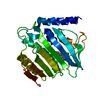
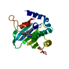
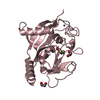
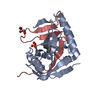
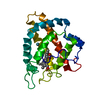
- PDB-1m55: Catalytic domain of the Adeno Associated Virus type 5 Rep protein -
+ Open data

- Basic information
| Entry | Database: PDB / ID: 1m55 | ||||||
|---|---|---|---|---|---|---|---|
| Title | Catalytic domain of the Adeno Associated Virus type 5 Rep protein | ||||||
 Components Components | Rep protein | ||||||
 Keywords Keywords | VIRAL PROTEIN / ENDONUCLEASE / REP / ROLLING CIRCLE REPLICATION / ADENO-ASSOCIATED VIRUS | ||||||
| Function / homology |  Function and homology information Function and homology informationviral genome replication / endonuclease activity / DNA replication / hydrolase activity / host cell nucleus / DNA binding / ATP binding / metal ion binding Similarity search - Function | ||||||
| Biological species |  Adeno-associated virus - 5 Adeno-associated virus - 5 | ||||||
| Method |  X-RAY DIFFRACTION / SYNCHROTRON / MAD / Resolution: 1.4 Å X-RAY DIFFRACTION / SYNCHROTRON / MAD / Resolution: 1.4 Å | ||||||
 Authors Authors | Hickman, A.B. / Ronning, D.R. / Kotin, R.M. / Dyda, F. | ||||||
 Citation Citation | Journal: Mol.Cell / Year: 2002 Title: Structural unity among viral origin binding proteins: crystal structure of the nuclease domain of adeno-associated virus Rep. Authors: Hickman, A.B. / Ronning, D.R. / Kotin, R.M. / Dyda, F. | ||||||
| History |
|
- Structure visualization
| Structure viewer | Molecule: MolmilJmol/JSmol |
|---|
- Downloads & links
-Download
| PDBx/mmCIF format | 1m55.cif.gz | 105.9 KB | Display | PDBx/mmCIF format |
|---|---|---|---|---|
| PDB format | pdb1m55.ent.gz | 80.1 KB | Display | PDB format |
| PDBx/mmJSON format | 1m55.json.gz | Tree view | PDBx/mmJSON format | |
| Others |  Other downloads Other downloads |
-Validation report
| Arichive directory | https://data.pdbj.org/pub/pdb/validation_reports/m5/1m55ftp://data.pdbj.org/pub/pdb/validation_reports/m5/1m55 | HTTPS FTP |
|---|
-Related structure data
| Similar structure data |
|---|


-Links
 PDBj
PDBj

- Assembly
| Deposited unit | 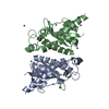
| ||||||||
|---|---|---|---|---|---|---|---|---|---|
| 1 | 
| ||||||||
| 2 | 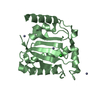
| ||||||||
| Unit cell |
|
-Components
| #1: Protein | Mass: 22776.982 Da / Num. of mol.: 2 / Fragment: Catalytic domain (Residues 1-197) Source method: isolated from a genetically manipulated source Source: (gene. exp.) Adeno-associated virus - 5 / Genus: Dependovirus / Plasmid: pET15b / Species (production host): Escherichia coli / Production host:  #2: Chemical | ChemComp-ZN /   Mass: 65.409 Da / Num. of mol.: 6 / Source method: obtained synthetically / Formula: Zn Mass: 65.409 Da / Num. of mol.: 6 / Source method: obtained synthetically / Formula: Zn#3: Chemical |   Mass: 35.453 Da / Num. of mol.: 2 / Source method: obtained synthetically / Formula: Cl Mass: 35.453 Da / Num. of mol.: 2 / Source method: obtained synthetically / Formula: Cl#4: Water | ChemComp-HOH / |  Mass: 18.015 Da / Num. of mol.: 675 / Source method: isolated from a natural source / Formula: H2O Mass: 18.015 Da / Num. of mol.: 675 / Source method: isolated from a natural source / Formula: H2O |
|---|
-Experimental details
-Experiment
| Experiment | Method: X-RAY DIFFRACTION / Number of used crystals: 1 |
|---|
- Sample preparation
| Crystal | Density Matthews: 2.26 Å3/Da / Density % sol: 45.55 % | ||||||||||||||||||||||||||||||||||||||||||||||||
|---|---|---|---|---|---|---|---|---|---|---|---|---|---|---|---|---|---|---|---|---|---|---|---|---|---|---|---|---|---|---|---|---|---|---|---|---|---|---|---|---|---|---|---|---|---|---|---|---|---|
| Crystal grow | Temperature: 293 K / Method: vapor diffusion, hanging drop / pH: 6.5 Details: PEG 8000, PEG 400, ZINC ACETATE, SODIUM CACODYLATE, TRIS, pH 6.5, VAPOR DIFFUSION, HANGING DROP, temperature 293K | ||||||||||||||||||||||||||||||||||||||||||||||||
| Crystal grow | *PLUS Temperature: 20 ℃ | ||||||||||||||||||||||||||||||||||||||||||||||||
| Components of the solutions | *PLUS
|
-Data collection
| Diffraction | Mean temperature: 95 K |
|---|---|
| Diffraction source | Source: SYNCHROTRON / Site: APS  / Beamline: 19-ID / Beamline: 19-ID |
| Detector | Type: CUSTOM-MADE / Detector: CCD / Date: Dec 6, 2001 |
| Radiation | Monochromator: Si(220) / Protocol: MAD / Monochromatic (M) / Laue (L): M / Scattering type: x-ray |
| Radiation wavelength | Relative weight: 1 |
| Reflection | Resolution: 1.4→30 Å / Num. all: 79390 / Num. obs: 79390 / % possible obs: 99.6 % / Observed criterion σ(F): 0 / Observed criterion σ(I): -3 / Redundancy: 4.71 % / Biso Wilson estimate: 13.1 Å2 / Rmerge(I) obs: 0.062 / Rsym value: 0.062 / Net I/σ(I): 10 |
| Reflection shell | Resolution: 1.4→1.43 Å / Redundancy: 2.68 % / Rmerge(I) obs: 0.404 / Mean I/σ(I) obs: 2.68 / Num. unique all: 4502 / Rsym value: 0.404 / % possible all: 95.9 |
| Reflection | *PLUS Num. measured all: 373647 / Rmerge(I) obs: 0.062 |
| Reflection shell | *PLUS % possible obs: 95.9 % / Rmerge(I) obs: 0.404 |
- Processing
| Software |
| ||||||||||||||||||||
|---|---|---|---|---|---|---|---|---|---|---|---|---|---|---|---|---|---|---|---|---|---|
| Refinement | Method to determine structure: MAD / Resolution: 1.4→30 Å / Cross valid method: THROUGHOUT / σ(F): 0 / Stereochemistry target values: Engh & Huber
| ||||||||||||||||||||
| Refinement step | Cycle: LAST / Resolution: 1.4→30 Å
| ||||||||||||||||||||
| Refine LS restraints |
| ||||||||||||||||||||
| LS refinement shell | Resolution: 1.4→1.46 Å / Rfactor Rfree error: 0.023
| ||||||||||||||||||||
| Refinement | *PLUS Highest resolution: 1.4 Å / Lowest resolution: 30 Å / Rfactor Rfree: 0.196 / Rfactor Rwork: 0.179 | ||||||||||||||||||||
| Solvent computation | *PLUS | ||||||||||||||||||||
| Displacement parameters | *PLUS | ||||||||||||||||||||
| Refine LS restraints | *PLUS Type: x_angle_deg / Dev ideal: 1.36 | ||||||||||||||||||||
| LS refinement shell | *PLUS Rfactor Rfree: 0.279 / Rfactor Rwork: 0.261 |
