 Movie
Movie Controller


+ Open data

- Basic information
| Entry | Database: PDB / ID: 3zjr | ||||||
|---|---|---|---|---|---|---|---|
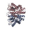
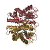
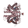
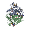
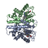
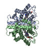
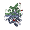
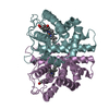
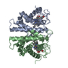
| Title | M.acetivorans protoglobin in complex with cyanide and Xenon | ||||||
 Components Components | PROTOGLOBIN | ||||||
 Keywords Keywords | IRON-BINDING PROTEIN / CYANIDE-XENON COMPLEX | ||||||
| Function / homology |  Function and homology information Function and homology information | ||||||
| Biological species |  METHANOSARCINA ACETIVORANS (archaea) METHANOSARCINA ACETIVORANS (archaea) | ||||||
| Method |  X-RAY DIFFRACTION / SYNCHROTRON / MOLECULAR REPLACEMENT / Resolution: 3 Å X-RAY DIFFRACTION / SYNCHROTRON / MOLECULAR REPLACEMENT / Resolution: 3 Å | ||||||
 Authors Authors | Pesce, A. / Tilleman, L. / Donne, J. / Aste, E. / Ascenzi, P. / Ciaccio, C. / Coletta, M. / Moens, L. / Viappiani, C. / Dewilde, S. ...Pesce, A. / Tilleman, L. / Donne, J. / Aste, E. / Ascenzi, P. / Ciaccio, C. / Coletta, M. / Moens, L. / Viappiani, C. / Dewilde, S. / Bolognesi, M. / Nardini, M. | ||||||
 Citation Citation | Journal: Plos One / Year: 2013 Title: Structure and Haem-Distal Site Plasticity in Methanosarcina Acetivorans Protoglobin. Authors: Pesce, A. / Tilleman, L. / Donne, J. / Aste, E. / Ascenzi, P. / Ciaccio, C. / Coletta, M. / Moens, L. / Viappiani, C. / Dewilde, S. / Bolognesi, M. / Nardini, M. | ||||||
| History |
|
- Structure visualization
| Structure viewer | Molecule: MolmilJmol/JSmol |
|---|
- Downloads & links
-Download
| PDBx/mmCIF format | 3zjr.cif.gz | 56 KB | Display | PDBx/mmCIF format |
|---|---|---|---|---|
| PDB format | pdb3zjr.ent.gz | 39.3 KB | Display | PDB format |
| PDBx/mmJSON format | 3zjr.json.gz | Tree view | PDBx/mmJSON format | |
| Others |  Other downloads Other downloads |
-Validation report
| Arichive directory | https://data.pdbj.org/pub/pdb/validation_reports/zj/3zjrftp://data.pdbj.org/pub/pdb/validation_reports/zj/3zjr | HTTPS FTP |
|---|
-Related structure data
| Related structure data |  3zjhC  3zjiC  3zjjC  3zjlC  3zjmC  3zjnC  3zjoC  3zjpC 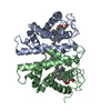 3zjqC  3zjsC  2vebS C: citing same article ( S: Starting model for refinement |
|---|---|
| Similar structure data |

-Links
 PDBj
PDBj


- Assembly
| Deposited unit | 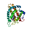
| ||||||||
|---|---|---|---|---|---|---|---|---|---|
| 1 | 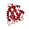
| ||||||||
| Unit cell |
|
-Components
| #1: Protein | Mass: 23010.863 Da / Num. of mol.: 1 / Mutation: YES Source method: isolated from a genetically manipulated source Source: (gene. exp.) METHANOSARCINA ACETIVORANS (archaea) / Production host:  |
|---|---|
| #2: Chemical | ChemComp-HEM /   Mass: 616.487 Da / Num. of mol.: 1 / Source method: obtained synthetically / Formula: C34H32FeN4O4 Mass: 616.487 Da / Num. of mol.: 1 / Source method: obtained synthetically / Formula: C34H32FeN4O4 |
| #3: Chemical | ChemComp-CYN /   Mass: 26.017 Da / Num. of mol.: 1 / Source method: obtained synthetically / Formula: CN Mass: 26.017 Da / Num. of mol.: 1 / Source method: obtained synthetically / Formula: CN |
| #4: Chemical | ChemComp-XE /   Mass: 131.293 Da / Num. of mol.: 1 / Source method: obtained synthetically / Formula: Xe Mass: 131.293 Da / Num. of mol.: 1 / Source method: obtained synthetically / Formula: Xe |
| #5: Water | ChemComp-HOH /  Mass: 18.015 Da / Num. of mol.: 22 / Source method: isolated from a natural source / Formula: H2O Mass: 18.015 Da / Num. of mol.: 22 / Source method: isolated from a natural source / Formula: H2O |
| Sequence details | C101S MUTATED FOR CRYSTALLIZ |
-Experimental details
-Experiment
| Experiment | Method: X-RAY DIFFRACTION / Number of used crystals: 1 |
|---|
- Sample preparation
| Crystal | Density Matthews: 2.22 Å3/Da / Density % sol: 44.56 % / Description: NONE |
|---|---|
| Crystal grow | pH: 7 Details: 20% PEG 4000, 10% ISOPROPANOL, 0.1 M HEPES PH 7.0, 0.02 M POTASSIUM FERRICYANIDE, 0.01 M KCN |
-Data collection
| Diffraction | Mean temperature: 100 K |
|---|---|
| Diffraction source | Source: SYNCHROTRON / Site: ESRF  / Beamline: ID14-1 / Wavelength: 1 / Beamline: ID14-1 / Wavelength: 1 |
| Detector | Type: ADSC CCD / Detector: CCD |
| Radiation | Protocol: SINGLE WAVELENGTH / Monochromatic (M) / Laue (L): M / Scattering type: x-ray |
| Radiation wavelength | Wavelength: 1 Å / Relative weight: 1 |
| Reflection | Resolution: 3→50.64 Å / Num. obs: 3627 / % possible obs: 87.8 % / Observed criterion σ(I): 1 / Redundancy: 1.9 % / Rmerge(I) obs: 0.14 / Net I/σ(I): 6.4 |
| Reflection shell | Resolution: 3→3.16 Å / Redundancy: 1.9 % / Rmerge(I) obs: 0.3 / Mean I/σ(I) obs: 2.3 / % possible all: 91.3 |
- Processing
| Software |
| ||||||||||||||||||||||||||||||||||||||||||||||||||||||||||||||||||||||||||||||||||||||||||||||||||||||||||||||||||||||||||||||||||||||||||||||||||||||||||||||||||||||||||||||||||||||
|---|---|---|---|---|---|---|---|---|---|---|---|---|---|---|---|---|---|---|---|---|---|---|---|---|---|---|---|---|---|---|---|---|---|---|---|---|---|---|---|---|---|---|---|---|---|---|---|---|---|---|---|---|---|---|---|---|---|---|---|---|---|---|---|---|---|---|---|---|---|---|---|---|---|---|---|---|---|---|---|---|---|---|---|---|---|---|---|---|---|---|---|---|---|---|---|---|---|---|---|---|---|---|---|---|---|---|---|---|---|---|---|---|---|---|---|---|---|---|---|---|---|---|---|---|---|---|---|---|---|---|---|---|---|---|---|---|---|---|---|---|---|---|---|---|---|---|---|---|---|---|---|---|---|---|---|---|---|---|---|---|---|---|---|---|---|---|---|---|---|---|---|---|---|---|---|---|---|---|---|---|---|---|---|
| Refinement | Method to determine structure: MOLECULAR REPLACEMENT Starting model: PDB ENTRY 2VEB Resolution: 3→42.37 Å / Cor.coef. Fo:Fc: 0.936 / Cor.coef. Fo:Fc free: 0.862 / SU B: 25.319 / SU ML: 0.433 / Cross valid method: THROUGHOUT / ESU R Free: 0.679 / Stereochemistry target values: MAXIMUM LIKELIHOOD Details: HYDROGENS HAVE BEEN ADDED IN THE RIDING POSITIONS. FINAL STRUCTURE HAS NO RESIDUES IN THE DISALLOWED REGION OF RAMACHANDRAN PLOT AS DEFINED IN THE CCP4 PROCHECK PROGRAM.
| ||||||||||||||||||||||||||||||||||||||||||||||||||||||||||||||||||||||||||||||||||||||||||||||||||||||||||||||||||||||||||||||||||||||||||||||||||||||||||||||||||||||||||||||||||||||
| Solvent computation | Ion probe radii: 0.8 Å / Shrinkage radii: 0.8 Å / VDW probe radii: 1.4 Å / Solvent model: MASK | ||||||||||||||||||||||||||||||||||||||||||||||||||||||||||||||||||||||||||||||||||||||||||||||||||||||||||||||||||||||||||||||||||||||||||||||||||||||||||||||||||||||||||||||||||||||
| Displacement parameters | Biso mean: 53.786 Å2
| ||||||||||||||||||||||||||||||||||||||||||||||||||||||||||||||||||||||||||||||||||||||||||||||||||||||||||||||||||||||||||||||||||||||||||||||||||||||||||||||||||||||||||||||||||||||
| Refinement step | Cycle: LAST / Resolution: 3→42.37 Å
| ||||||||||||||||||||||||||||||||||||||||||||||||||||||||||||||||||||||||||||||||||||||||||||||||||||||||||||||||||||||||||||||||||||||||||||||||||||||||||||||||||||||||||||||||||||||
| Refine LS restraints |
|