 Movie
Movie Controller

[English] 日本語
 Yorodumi
- PDB-3ls4: Crystal Structure of Anti-tetrahydrocannabinol Fab Fragment in Co... -
+ Open data

- Basic information
| Entry | Database: PDB / ID: 3ls4 | ||||||
|---|---|---|---|---|---|---|---|
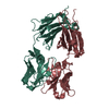
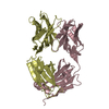
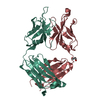
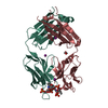
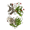
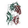
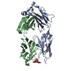
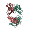
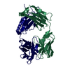
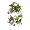
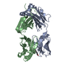
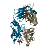
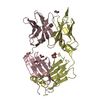
| Title | Crystal Structure of Anti-tetrahydrocannabinol Fab Fragment in Complex with THC | ||||||
 Components Components |
| ||||||
 Keywords Keywords | IMMUNE SYSTEM / Antibody / Immunoglobulin / Fab fragment / Cannabinoid complex | ||||||
| Function / homology | Immunoglobulins / Immunoglobulin-like / Sandwich / Mainly Beta / Chem-TCI Function and homology information Function and homology information | ||||||
| Biological species |  | ||||||
| Method |  X-RAY DIFFRACTION / SYNCHROTRON / MOLECULAR REPLACEMENT / Resolution: 2 Å X-RAY DIFFRACTION / SYNCHROTRON / MOLECULAR REPLACEMENT / Resolution: 2 Å | ||||||
 Authors Authors | Niemi, M.H. / Rouvinen, J. | ||||||
 Citation Citation | Journal: J.Mol.Biol. / Year: 2010 Title: A structural insight into the molecular recognition of a (-)-Delta9-tetrahydrocannabinol and the development of a sensitive, one-step, homogeneous immunocomplex-based assay for its detection Authors: Niemi, M.H. / Turunen, L. / Pulli, T. / Nevanen, T.K. / Hoyhtya, M. / Soderlund, H. / Rouvinen, J. / Takkinen, K. | ||||||
| History |
|
- Structure visualization
| Structure viewer | Molecule: MolmilJmol/JSmol |
|---|
- Downloads & links
-Download
| PDBx/mmCIF format | 3ls4.cif.gz | 101.9 KB | Display | PDBx/mmCIF format |
|---|---|---|---|---|
| PDB format | pdb3ls4.ent.gz | 77.3 KB | Display | PDB format |
| PDBx/mmJSON format | 3ls4.json.gz | Tree view | PDBx/mmJSON format | |
| Others |  Other downloads Other downloads |
-Validation report
| Arichive directory | https://data.pdbj.org/pub/pdb/validation_reports/ls/3ls4ftp://data.pdbj.org/pub/pdb/validation_reports/ls/3ls4 | HTTPS FTP |
|---|
-Related structure data


-Links
 PDBj
PDBj

- Assembly
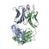
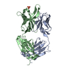
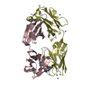
| Deposited unit | 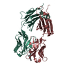
| ||||||||
|---|---|---|---|---|---|---|---|---|---|
| 1 |
| ||||||||
| Unit cell |
|
-Components
| #1: Antibody | Mass: 23346.822 Da / Num. of mol.: 1 Source method: isolated from a genetically manipulated source Source: (gene. exp.)  |
|---|---|
| #2: Antibody | Mass: 23371.443 Da / Num. of mol.: 1 Source method: isolated from a genetically manipulated source Source: (gene. exp.) |
| #3: Chemical | ChemComp-TCI / (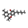  Mass: 314.462 Da / Num. of mol.: 1 / Source method: obtained synthetically / Formula: C21H30O2 Mass: 314.462 Da / Num. of mol.: 1 / Source method: obtained synthetically / Formula: C21H30O2 |
| #4: Water | ChemComp-HOH /  Mass: 18.015 Da / Num. of mol.: 240 / Source method: isolated from a natural source / Formula: H2O Mass: 18.015 Da / Num. of mol.: 240 / Source method: isolated from a natural source / Formula: H2O |
| Has protein modification | Y |
-Experimental details
-Experiment
| Experiment | Method: X-RAY DIFFRACTION / Number of used crystals: 1 |
|---|
- Sample preparation
| Crystal | Density Matthews: 2.96 Å3/Da / Density % sol: 58.39 % |
|---|---|
| Crystal grow | Temperature: 293 K / Method: vapor diffusion, hanging drop / pH: 7.5 Details: 12% PEG 3350, 0.1M sodium Hepes, pH 7.5, VAPOR DIFFUSION, HANGING DROP, temperature 293K |
-Data collection
| Diffraction | Mean temperature: 100 K |
|---|---|
| Diffraction source | Source: SYNCHROTRON / Site: ESRF  / Beamline: ID14-4 / Wavelength: 0.9395 Å / Beamline: ID14-4 / Wavelength: 0.9395 Å |
| Detector | Type: ADSC QUANTUM 315r / Detector: CCD / Date: Jan 30, 2009 |
| Radiation | Monochromator: Si (111) single crystal / Protocol: SINGLE WAVELENGTH / Monochromatic (M) / Laue (L): M / Scattering type: x-ray |
| Radiation wavelength | Wavelength: 0.9395 Å / Relative weight: 1 |
| Reflection | Resolution: 2→50 Å / Num. all: 38385 / Num. obs: 38070 / % possible obs: 99.2 % / Observed criterion σ(F): 2 / Observed criterion σ(I): 2 / Redundancy: 7.1 % / Biso Wilson estimate: 36.28 Å2 / Rsym value: 0.039 |
| Reflection shell | Resolution: 2→2.1 Å / Redundancy: 7.3 % / Mean I/σ(I) obs: 4.5 / Num. unique all: 5138 / Rsym value: 0.445 / % possible all: 99.9 |
- Processing
| Software |
| |||||||||||||||||||||||||||||||||||||||||||||||||||||||||||||||||||||||||||||||||||||||||||||||||||||||||
|---|---|---|---|---|---|---|---|---|---|---|---|---|---|---|---|---|---|---|---|---|---|---|---|---|---|---|---|---|---|---|---|---|---|---|---|---|---|---|---|---|---|---|---|---|---|---|---|---|---|---|---|---|---|---|---|---|---|---|---|---|---|---|---|---|---|---|---|---|---|---|---|---|---|---|---|---|---|---|---|---|---|---|---|---|---|---|---|---|---|---|---|---|---|---|---|---|---|---|---|---|---|---|---|---|---|---|
| Refinement | Method to determine structure: MOLECULAR REPLACEMENT Starting model: Free form structure of the same antibody Resolution: 2→35.731 Å / Occupancy max: 1 / Occupancy min: 0.39 / FOM work R set: 0.791 / SU ML: 0.32 / σ(F): 2 / Stereochemistry target values: ML
| |||||||||||||||||||||||||||||||||||||||||||||||||||||||||||||||||||||||||||||||||||||||||||||||||||||||||
| Solvent computation | Shrinkage radii: 0.9 Å / VDW probe radii: 1.11 Å / Solvent model: FLAT BULK SOLVENT MODEL / Bsol: 40.962 Å2 / ksol: 0.337 e/Å3 | |||||||||||||||||||||||||||||||||||||||||||||||||||||||||||||||||||||||||||||||||||||||||||||||||||||||||
| Displacement parameters | Biso max: 79.43 Å2 / Biso mean: 39.22 Å2 / Biso min: 19.04 Å2
| |||||||||||||||||||||||||||||||||||||||||||||||||||||||||||||||||||||||||||||||||||||||||||||||||||||||||
| Refinement step | Cycle: LAST / Resolution: 2→35.731 Å
| |||||||||||||||||||||||||||||||||||||||||||||||||||||||||||||||||||||||||||||||||||||||||||||||||||||||||
| Refine LS restraints |
| |||||||||||||||||||||||||||||||||||||||||||||||||||||||||||||||||||||||||||||||||||||||||||||||||||||||||
| LS refinement shell | Refine-ID: X-RAY DIFFRACTION / Total num. of bins used: 14
|
