+ Open data
- Basic information
| Entry | Database: PDB / ID: 3hl2 | ||||||
|---|---|---|---|---|---|---|---|
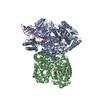
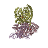
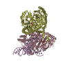
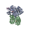
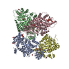
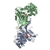
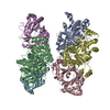
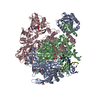
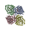
| Title | The crystal structure of the human SepSecS-tRNASec complex | ||||||
 Components Components |
| ||||||
 Keywords Keywords | TRANSFERASE / selenocysteine / tRNASec / SepSecS / protein-RNA complex / Alternative splicing / Cytoplasm / Protein biosynthesis / Pyridoxal phosphate / Selenium | ||||||
| Function / homology |  Function and homology information Function and homology informationO-phospho-L-seryl-tRNASec:L-selenocysteinyl-tRNA synthase / O-phosphoseryl-tRNA(Sec) selenium transferase activity / conversion of seryl-tRNAsec to selenocys-tRNAsec / selenocysteine incorporation / Selenocysteine synthesis / tRNA binding / nucleus / cytosol / cytoplasm Similarity search - Function | ||||||
| Biological species |  Homo sapiens (human) Homo sapiens (human) | ||||||
| Method |  X-RAY DIFFRACTION / X-RAY DIFFRACTION /  SYNCHROTRON / SYNCHROTRON /  MOLECULAR REPLACEMENT / Resolution: 2.81 Å MOLECULAR REPLACEMENT / Resolution: 2.81 Å | ||||||
 Authors Authors | Palioura, S. / Steitz, T.A. / Soll, D. / Simonovic, M. | ||||||
 Citation Citation |  Journal: Science / Year: 2009 Journal: Science / Year: 2009Title: The human SepSecS-tRNASec complex reveals the mechanism of selenocysteine formation. Authors: Palioura, S. / Sherrer, R.L. / Steitz, T.A. / Soll, D. / Simonovic, M. | ||||||
| History |
|
- Structure visualization
| Structure viewer | Molecule:  Molmil Molmil Jmol/JSmol Jmol/JSmol |
|---|
- Downloads & links
- Download
| PDBx/mmCIF format |  3hl2.cif.gz 3hl2.cif.gz | 436.8 KB | Display |  PDBx/mmCIF format PDBx/mmCIF format |
|---|---|---|---|---|
| PDB format |  pdb3hl2.ent.gz pdb3hl2.ent.gz | 356.5 KB | Display |  PDB format PDB format |
| PDBx/mmJSON format |  3hl2.json.gz 3hl2.json.gz | Tree view |  PDBx/mmJSON format PDBx/mmJSON format | |
| Others |  Other downloads Other downloads |
-Validation report
| Summary document |  3hl2_validation.pdf.gz 3hl2_validation.pdf.gz | 521.3 KB | Display |  wwPDB validaton report wwPDB validaton report |
|---|---|---|---|---|
| Full document |  3hl2_full_validation.pdf.gz 3hl2_full_validation.pdf.gz | 580.8 KB | Display | |
| Data in XML |  3hl2_validation.xml.gz 3hl2_validation.xml.gz | 73.3 KB | Display | |
| Data in CIF |  3hl2_validation.cif.gz 3hl2_validation.cif.gz | 102.6 KB | Display | |
| Arichive directory |  https://data.pdbj.org/pub/pdb/validation_reports/hl/3hl2 https://data.pdbj.org/pub/pdb/validation_reports/hl/3hl2 ftp://data.pdbj.org/pub/pdb/validation_reports/hl/3hl2 ftp://data.pdbj.org/pub/pdb/validation_reports/hl/3hl2 | HTTPS FTP |
-Related structure data
| Related structure data | 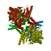 3bc8S S: Starting model for refinement |
|---|---|
| Similar structure data |
- Links
Links
- Assembly
| Deposited unit | 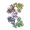
| |||||||||
|---|---|---|---|---|---|---|---|---|---|---|
| 1 | 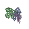
| |||||||||
| 2 | 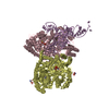
| |||||||||
| Unit cell |
| |||||||||
| Components on special symmetry positions |
| |||||||||
| Details | The physiologically active tetrameric assembly can be reconstructed by applying the following symmetry operation onto the chains A and B, and the conformer A of the chain E: X, X-Y, -Z (0 -1 0). Similarly, the physiologic tetramer can be built by applying the symmetry operation onto the chains C and D, and the conformer B of the chain E: -X+Y, Y, -Z+1/3 (0 0 0). |
- Components
-Protein / RNA chain , 2 types, 5 molecules ABCDE
| #1: Protein | Mass: 55801.211 Da / Num. of mol.: 4 Source method: isolated from a genetically manipulated source Source: (gene. exp.)  Homo sapiens (human) / Gene: SEPSECS, TRNP48 / Plasmid: pET15b / Production host: Homo sapiens (human) / Gene: SEPSECS, TRNP48 / Plasmid: pET15b / Production host:  References: UniProt: Q9HD40, L-seryl-tRNASec selenium transferase #2: RNA chain | | Mass: 28908.084 Da / Num. of mol.: 1 Source method: isolated from a genetically manipulated source Source: (gene. exp.)  Homo sapiens (human) / Plasmid: pUC19 / Production host: Homo sapiens (human) / Plasmid: pUC19 / Production host:  |
|---|
-Non-polymers , 4 types, 299 molecules 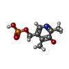
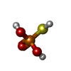
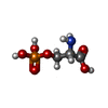


| #3: Chemical | ChemComp-PLR / ( #4: Chemical | #5: Chemical | ChemComp-SEP / #6: Water | ChemComp-HOH / | |
|---|
-Details
| Has protein modification | Y |
|---|
-Experimental details
-Experiment
| Experiment | Method:  X-RAY DIFFRACTION / Number of used crystals: 1 X-RAY DIFFRACTION / Number of used crystals: 1 |
|---|
- Sample preparation
| Crystal | Density Matthews: 3.77 Å3/Da / Density % sol: 67.33 % |
|---|---|
| Crystal grow | Temperature: 285 K / pH: 6.5 Details: 0.3M tri-lithium citrate, 18% (w/v) PEG 3,350, pH 6.5, VAPOR DIFFUSION, SITTING DROP, temperature 285K |
-Data collection
| Diffraction | Mean temperature: 77 K |
|---|---|
| Diffraction source | Source:  SYNCHROTRON / Site: SYNCHROTRON / Site:  APS APS  / Beamline: 24-ID-E / Wavelength: 0.97918 / Beamline: 24-ID-E / Wavelength: 0.97918 |
| Detector | Type: ADSC QUANTUM 315 / Detector: CCD / Date: Aug 10, 2008 |
| Radiation | Monochromator: SAGITALLY FOCUSED SI(111) / Protocol: SINGLE WAVELENGTH / Monochromatic (M) / Laue (L): M / Scattering type: x-ray |
| Radiation wavelength | Wavelength: 0.97918 Å / Relative weight: 1 |
| Reflection | Resolution: 2.8→50 Å / Num. obs: 89375 / % possible obs: 97.5 % / Observed criterion σ(I): 0 / Redundancy: 5.4 % / Biso Wilson estimate: 65.3 Å2 / Rsym value: 0.17 / Net I/σ(I): 6 |
| Reflection shell | Resolution: 2.8→2.9 Å / Redundancy: 4.3 % / Mean I/σ(I) obs: 1 / Rsym value: 0.94 / % possible all: 94.9 |
- Processing
| Software |
| |||||||||||||||||||||||||||||||||||||||||||||||||||||||||||||||||||||||||||||||||||||||||||||||||||||||||
|---|---|---|---|---|---|---|---|---|---|---|---|---|---|---|---|---|---|---|---|---|---|---|---|---|---|---|---|---|---|---|---|---|---|---|---|---|---|---|---|---|---|---|---|---|---|---|---|---|---|---|---|---|---|---|---|---|---|---|---|---|---|---|---|---|---|---|---|---|---|---|---|---|---|---|---|---|---|---|---|---|---|---|---|---|---|---|---|---|---|---|---|---|---|---|---|---|---|---|---|---|---|---|---|---|---|---|
| Refinement | Method to determine structure:  MOLECULAR REPLACEMENT MOLECULAR REPLACEMENTStarting model: 3BC8 Resolution: 2.81→37.95 Å / SU ML: 0.4 / σ(F): 1.34 / Stereochemistry target values: ML
| |||||||||||||||||||||||||||||||||||||||||||||||||||||||||||||||||||||||||||||||||||||||||||||||||||||||||
| Solvent computation | Shrinkage radii: 0.9 Å / VDW probe radii: 1.11 Å / Solvent model: FLAT BULK SOLVENT MODEL / Bsol: 48.94 Å2 / ksol: 0.28 e/Å3 | |||||||||||||||||||||||||||||||||||||||||||||||||||||||||||||||||||||||||||||||||||||||||||||||||||||||||
| Refinement step | Cycle: LAST / Resolution: 2.81→37.95 Å
| |||||||||||||||||||||||||||||||||||||||||||||||||||||||||||||||||||||||||||||||||||||||||||||||||||||||||
| LS refinement shell |
|
 Movie
Movie Controller


 PDBj
PDBj


