 Movie
Movie Controller

[English] 日本語
 Yorodumi
- PDB-3emw: Crystal Structure of human splA/ryanodine receptor domain and SOC... -
+ Open data

- Basic information
| Entry | Database: PDB / ID: 3emw | ||||||
|---|---|---|---|---|---|---|---|
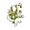
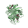
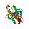
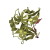
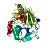
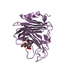
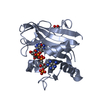
| Title | Crystal Structure of human splA/ryanodine receptor domain and SOCS box containing 2 (SPSB2) in complex with a 20-residue VASA peptide | ||||||
 Components Components |
| ||||||
 Keywords Keywords | APOPTOSIS / Apoptosis nucleus / UBL conjugation pathwayc / CL transcription regulation / Transcription / Phosphoprotein / Ubl conjugation pathway / Structural Genomics / Structural Genomics Consortium / SGC | ||||||
| Function / homology |  Function and homology information Function and homology informationposterior cell cortex / secondary piRNA processing / gamete generation / P granule / germ cell nucleus / SCF ubiquitin ligase complex / oogenesis / germ cell development / ubiquitin-like ligase-substrate adaptor activity / translation initiation factor binding ...posterior cell cortex / secondary piRNA processing / gamete generation / P granule / germ cell nucleus / SCF ubiquitin ligase complex / oogenesis / germ cell development / ubiquitin-like ligase-substrate adaptor activity / translation initiation factor binding / intracellular protein localization / Antigen processing: Ubiquitination & Proteasome degradation / Neddylation / ubiquitin-dependent protein catabolic process / proteasome-mediated ubiquitin-dependent protein catabolic process / cell differentiation / RNA helicase activity / intracellular signal transduction / RNA helicase / protein ubiquitination / mRNA binding / perinuclear region of cytoplasm / ATP hydrolysis activity / ATP binding / nucleus / cytoplasm / cytosol Similarity search - Function | ||||||
| Biological species |  Homo sapiens (human) Homo sapiens (human) | ||||||
| Method |  X-RAY DIFFRACTION / SYNCHROTRON / MOLECULAR REPLACEMENT / Resolution: 1.8 Å X-RAY DIFFRACTION / SYNCHROTRON / MOLECULAR REPLACEMENT / Resolution: 1.8 Å | ||||||
 Authors Authors | Filippakopoulos, P. / Sharpe, T. / Keates, T. / Murray, J.W. / Savitsky, P. / Roos, A.K. / Pike, A.C.W. / Von Delft, F. / Arrowsmith, C.H. / Edwards, A.M. ...Filippakopoulos, P. / Sharpe, T. / Keates, T. / Murray, J.W. / Savitsky, P. / Roos, A.K. / Pike, A.C.W. / Von Delft, F. / Arrowsmith, C.H. / Edwards, A.M. / Weigelt, J. / Bountra, C. / Knapp, S. / Bullock, A. / Structural Genomics Consortium (SGC) | ||||||
 Citation Citation | Journal: J.Mol.Biol. / Year: 2010 Title: Structural basis for Par-4 recognition by the SPRY domain- and SOCS box-containing proteins SPSB1, SPSB2, and SPSB4. Authors: Filippakopoulos, P. / Low, A. / Sharpe, T.D. / Uppenberg, J. / Yao, S. / Kuang, Z. / Savitsky, P. / Lewis, R.S. / Nicholson, S.E. / Norton, R.S. / Bullock, A.N. | ||||||
| History |
|
- Structure visualization
| Structure viewer | Molecule: MolmilJmol/JSmol |
|---|
- Downloads & links
-Download
| PDBx/mmCIF format | 3emw.cif.gz | 58.4 KB | Display | PDBx/mmCIF format |
|---|---|---|---|---|
| PDB format | pdb3emw.ent.gz | 41.6 KB | Display | PDB format |
| PDBx/mmJSON format | 3emw.json.gz | Tree view | PDBx/mmJSON format | |
| Others |  Other downloads Other downloads |
-Validation report
| Arichive directory | https://data.pdbj.org/pub/pdb/validation_reports/em/3emwftp://data.pdbj.org/pub/pdb/validation_reports/em/3emw | HTTPS FTP |
|---|
-Related structure data
| Related structure data |  2jk9SC  2v24C  3f2oC S: Starting model for refinement C: citing same article ( |
|---|---|
| Similar structure data |


-Links
 PDBj
PDBj
- Assembly
| Deposited unit | 
| ||||||||
|---|---|---|---|---|---|---|---|---|---|
| 1 |
| ||||||||
| Unit cell |
|
-Components
| #1: Protein | Mass: 23849.627 Da / Num. of mol.: 1 / Fragment: UNP residues 26-219 Source method: isolated from a genetically manipulated source Source: (gene. exp.) Homo sapiens (human) / Gene: SPSB2, GRCC9, SSB2 / Plasmid: pNIC28-Bsa4 / Production host:  | ||
|---|---|---|---|
| #2: Protein/peptide | Mass: 2497.696 Da / Num. of mol.: 1 / Source method: obtained synthetically / Details: Peptide synthesis / References: UniProt: P09052*PLUS | ||
| #3: Chemical | ChemComp-EDO /   Mass: 62.068 Da / Num. of mol.: 4 / Source method: obtained synthetically / Formula: C2H6O2 Mass: 62.068 Da / Num. of mol.: 4 / Source method: obtained synthetically / Formula: C2H6O2#4: Water | ChemComp-HOH / |  Mass: 18.015 Da / Num. of mol.: 138 / Source method: isolated from a natural source / Formula: H2O Mass: 18.015 Da / Num. of mol.: 138 / Source method: isolated from a natural source / Formula: H2O |
-Experimental details
-Experiment
| Experiment | Method: X-RAY DIFFRACTION / Number of used crystals: 1 |
|---|
- Sample preparation
| Crystal | Density Matthews: 2.4 Å3/Da / Density % sol: 48.82 % |
|---|---|
| Crystal grow | Temperature: 277 K / Method: vapor diffusion, sitting drop / pH: 7.5 Details: 0.178M Na/KPO4, 17.8% PEG3350, 8.88% EtGly, pH7.5, VAPOR DIFFUSION, SITTING DROP, temperature 277K |
-Data collection
| Diffraction | Mean temperature: 100 K |
|---|---|
| Diffraction source | Source: SYNCHROTRON / Site: Diamond  / Beamline: I03 / Wavelength: 0.975653 Å / Beamline: I03 / Wavelength: 0.975653 Å |
| Detector | Type: ADSC QUANTUM 315 / Detector: CCD / Date: Sep 21, 2008 |
| Radiation | Protocol: SINGLE WAVELENGTH / Monochromatic (M) / Laue (L): M / Scattering type: x-ray |
| Radiation wavelength | Wavelength: 0.975653 Å / Relative weight: 1 |
| Reflection | Resolution: 1.8→42.83 Å / Num. obs: 24366 / % possible obs: 99.9 % / Redundancy: 6.9 % / Biso Wilson estimate: 25.22 Å2 / Rmerge(I) obs: 0.078 / Rsym value: 0.078 / Net I/σ(I): 15 |
| Reflection shell | Resolution: 1.8→1.9 Å / Redundancy: 7.1 % / Rmerge(I) obs: 0.618 / Mean I/σ(I) obs: 3.1 / Num. unique all: 3494 / Rsym value: 0.618 / % possible all: 100 |
- Processing
| Software |
| ||||||||||||||||||||||||||||||||||||||||||||||||||||||||||||||||||||||||||||||||||||||||||||||||||||
|---|---|---|---|---|---|---|---|---|---|---|---|---|---|---|---|---|---|---|---|---|---|---|---|---|---|---|---|---|---|---|---|---|---|---|---|---|---|---|---|---|---|---|---|---|---|---|---|---|---|---|---|---|---|---|---|---|---|---|---|---|---|---|---|---|---|---|---|---|---|---|---|---|---|---|---|---|---|---|---|---|---|---|---|---|---|---|---|---|---|---|---|---|---|---|---|---|---|---|---|---|---|
| Refinement | Method to determine structure: MOLECULAR REPLACEMENT Starting model: PDB ENTRY 2JK9 Resolution: 1.8→42.83 Å / Cor.coef. Fo:Fc: 0.96 / Cor.coef. Fo:Fc free: 0.945 / SU B: 4.491 / SU ML: 0.074 / TLS residual ADP flag: LIKELY RESIDUAL / Cross valid method: THROUGHOUT / ESU R: 0.107 / ESU R Free: 0.109 / Stereochemistry target values: MAXIMUM LIKELIHOOD / Details: HYDROGENS HAVE BEEN ADDED IN THE RIDING POSITIONS
| ||||||||||||||||||||||||||||||||||||||||||||||||||||||||||||||||||||||||||||||||||||||||||||||||||||
| Solvent computation | Ion probe radii: 0.8 Å / Shrinkage radii: 0.8 Å / VDW probe radii: 1.2 Å / Solvent model: MASK | ||||||||||||||||||||||||||||||||||||||||||||||||||||||||||||||||||||||||||||||||||||||||||||||||||||
| Displacement parameters | Biso mean: 18.523 Å2
| ||||||||||||||||||||||||||||||||||||||||||||||||||||||||||||||||||||||||||||||||||||||||||||||||||||
| Refinement step | Cycle: LAST / Resolution: 1.8→42.83 Å
| ||||||||||||||||||||||||||||||||||||||||||||||||||||||||||||||||||||||||||||||||||||||||||||||||||||
| Refine LS restraints |
| ||||||||||||||||||||||||||||||||||||||||||||||||||||||||||||||||||||||||||||||||||||||||||||||||||||
| LS refinement shell | Resolution: 1.8→1.847 Å / Total num. of bins used: 20
| ||||||||||||||||||||||||||||||||||||||||||||||||||||||||||||||||||||||||||||||||||||||||||||||||||||
| Refinement TLS params. | Method: refined / Refine-ID: X-RAY DIFFRACTION
| ||||||||||||||||||||||||||||||||||||||||||||||||||||||||||||||||||||||||||||||||||||||||||||||||||||
| Refinement TLS group |
|
