 Movie
Movie Controller

[English] 日本語
 Yorodumi
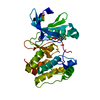
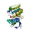
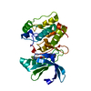
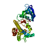
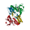
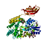
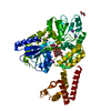
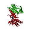
- PDB-3dj5: Crystal structure of the mouse Aurora-A catalytic domain (Asn186-... -
+ Open data

- Basic information
| Entry | Database: PDB / ID: 3dj5 | ||||||
|---|---|---|---|---|---|---|---|
| Title | Crystal structure of the mouse Aurora-A catalytic domain (Asn186->Gly, Lys240->Arg, Met302->Leu) in complex with Compound 290. | ||||||
 Components Components | serine/threonine kinase 6 | ||||||
 Keywords Keywords | TRANSFERASE / Aurora A / small-molecule inhibitor / fragment-based drug discovery / Kinase | ||||||
| Function / homology |  Function and homology information Function and homology informationInteraction between PHLDA1 and AURKA / TP53 Regulates Transcription of Genes Involved in G2 Cell Cycle Arrest / APC/C:Cdh1 mediated degradation of Cdc20 and other APC/C:Cdh1 targeted proteins in late mitosis/early G1 / FBXL7 down-regulates AURKA during mitotic entry and in early mitosis / AURKA Activation by TPX2 / meiotic spindle organization / Regulation of TP53 Activity through Phosphorylation / Regulation of PLK1 Activity at G2/M Transition / axon hillock / spindle assembly involved in female meiosis I ...Interaction between PHLDA1 and AURKA / TP53 Regulates Transcription of Genes Involved in G2 Cell Cycle Arrest / APC/C:Cdh1 mediated degradation of Cdc20 and other APC/C:Cdh1 targeted proteins in late mitosis/early G1 / FBXL7 down-regulates AURKA during mitotic entry and in early mitosis / AURKA Activation by TPX2 / meiotic spindle organization / Regulation of TP53 Activity through Phosphorylation / Regulation of PLK1 Activity at G2/M Transition / axon hillock / spindle assembly involved in female meiosis I / cilium disassembly / spindle pole centrosome / histone H3S10 kinase activity / chromosome passenger complex / positive regulation of oocyte maturation / mitotic centrosome separation / pronucleus / germinal vesicle / protein localization to centrosome / meiotic spindle / anterior/posterior axis specification / spindle organization / neuron projection extension / centrosome localization / centrosome cycle / positive regulation of mitochondrial fission / mitotic spindle pole / spindle midzone / microtubule organizing center / positive regulation of mitotic cell cycle / liver regeneration / molecular function activator activity / mitotic spindle organization / regulation of cytokinesis / spindle microtubule / meiotic cell cycle / centriole / regulation of protein stability / response to wounding / kinetochore / microtubule cytoskeleton organization / spindle pole / mitotic spindle / mitotic cell cycle / positive regulation of proteasomal ubiquitin-dependent protein catabolic process / microtubule cytoskeleton / basolateral plasma membrane / protein kinase activity / non-specific serine/threonine protein kinase / postsynaptic density / ciliary basal body / protein heterodimerization activity / negative regulation of gene expression / protein serine kinase activity / cell division / protein serine/threonine kinase activity / ubiquitin protein ligase binding / centrosome / protein kinase binding / negative regulation of apoptotic process / perinuclear region of cytoplasm / glutamatergic synapse / nucleoplasm / ATP binding / nucleus / cytosol Similarity search - Function | ||||||
| Biological species |  | ||||||
| Method |  X-RAY DIFFRACTION / SYNCHROTRON / MOLECULAR REPLACEMENT / Resolution: 1.8 Å X-RAY DIFFRACTION / SYNCHROTRON / MOLECULAR REPLACEMENT / Resolution: 1.8 Å | ||||||
 Authors Authors | Elling, R.A. / Erlanson, D.A. / Yang, W. / Tangonan, B.T. / Hansen, S.K. / Romanowski, M.J. | ||||||
 Citation Citation | Journal: To be Published Title: New fragment-based drug discovery Authors: Elling, R.A. / A Erlanson, D. / Yang, W. / Tangonan, B.T. / Hansen, S.K. / Romanowski, M.J. | ||||||
| History |
|
- Structure visualization
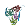
| Structure viewer | Molecule: MolmilJmol/JSmol |
|---|
- Downloads & links
-Download
| PDBx/mmCIF format | 3dj5.cif.gz | 76.8 KB | Display | PDBx/mmCIF format |
|---|---|---|---|---|
| PDB format | pdb3dj5.ent.gz | 55 KB | Display | PDB format |
| PDBx/mmJSON format | 3dj5.json.gz | Tree view | PDBx/mmJSON format | |
| Others |  Other downloads Other downloads |
-Validation report
| Arichive directory | https://data.pdbj.org/pub/pdb/validation_reports/dj/3dj5ftp://data.pdbj.org/pub/pdb/validation_reports/dj/3dj5 | HTTPS FTP |
|---|
-Related structure data
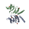
| Related structure data | 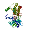 3dj6C 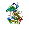 3dj7C 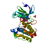 2c6dS S: Starting model for refinement C: citing same article ( |
|---|---|
| Similar structure data |

-Links
 PDBj
PDBj


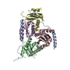
- Assembly
| Deposited unit | 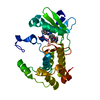
| |||||||||
|---|---|---|---|---|---|---|---|---|---|---|
| 1 | 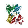
| |||||||||
| 2 |
| |||||||||
| Unit cell |
| |||||||||
| Components on special symmetry positions |
|
-Components
| #1: Protein | Mass: 31549.174 Da / Num. of mol.: 1 / Fragment: Aurora A kinase domain, UNP residues 116-382 / Mutation: N186G, K240R, M302L Source method: isolated from a genetically manipulated source Source: (gene. exp.)  References: UniProt: Q8C3H8, UniProt: P97477*PLUS, non-specific serine/threonine protein kinase |
|---|---|
| #2: Chemical | ChemComp-AK5 / 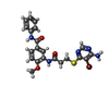  Mass: 502.384 Da / Num. of mol.: 1 / Source method: obtained synthetically / Formula: C21H20BrN5O3S Mass: 502.384 Da / Num. of mol.: 1 / Source method: obtained synthetically / Formula: C21H20BrN5O3S |
| #3: Water | ChemComp-HOH /  Mass: 18.015 Da / Num. of mol.: 369 / Source method: isolated from a natural source / Formula: H2O Mass: 18.015 Da / Num. of mol.: 369 / Source method: isolated from a natural source / Formula: H2O |
-Experimental details
-Experiment
| Experiment | Method: X-RAY DIFFRACTION / Number of used crystals: 1 |
|---|
- Sample preparation
| Crystal | Density Matthews: 2.72 Å3/Da / Density % sol: 54.77 % |
|---|---|
| Crystal grow | Temperature: 293 K / pH: 5.6 Details: Protein at 8.5 mg/ml in 50 mM Tris pH 7.0, 200 mM NaCl, 3 mM DTT; hanging-drop vapor diffusion; mother liquor: 0.1 M sodium citrate tribasic dihydrate pH 5.6, 2% tacsimate, 16% PEG 3350; ...Details: Protein at 8.5 mg/ml in 50 mM Tris pH 7.0, 200 mM NaCl, 3 mM DTT; hanging-drop vapor diffusion; mother liquor: 0.1 M sodium citrate tribasic dihydrate pH 5.6, 2% tacsimate, 16% PEG 3350; temperature: 293K; cryoprotectant: 20% glycerol; crystal frozen by immersion in liquid nitrogen |
-Data collection
| Diffraction | Mean temperature: 160 K |
|---|---|
| Diffraction source | Source: SYNCHROTRON / Site: SSRL  / Beamline: BL7-1 / Wavelength: 0.98 Å / Beamline: BL7-1 / Wavelength: 0.98 Å |
| Detector | Type: ADSC QUANTUM 315 / Detector: CCD / Date: Apr 29, 2008 / Details: Vertical focusing mirror |
| Radiation | Monochromator: single crystal (Si111) bent monochromator (horizontal focusing) Protocol: SINGLE WAVELENGTH / Monochromatic (M) / Laue (L): M / Scattering type: x-ray |
| Radiation wavelength | Wavelength: 0.98 Å / Relative weight: 1 |
| Reflection | Resolution: 1.8→30 Å / Num. all: 32098 / Num. obs: 31664 / % possible obs: 98.7 % / Redundancy: 3.8 % / Rmerge(I) obs: 0.053 / Net I/σ(I): 16.6 |
| Reflection shell | Resolution: 1.8→1.86 Å / Rmerge(I) obs: 0.167 / Mean I/σ(I) obs: 4.9 / % possible all: 99.8 |
- Processing
| Software |
| ||||||||||||||||||||||||||||||||||||||||||||||||||||||||||||||||||||||||||||||||||||||||||||||||||||||||||||||||||||||||||||||||||||||||||||||||||||||||||||||||||||||||||
|---|---|---|---|---|---|---|---|---|---|---|---|---|---|---|---|---|---|---|---|---|---|---|---|---|---|---|---|---|---|---|---|---|---|---|---|---|---|---|---|---|---|---|---|---|---|---|---|---|---|---|---|---|---|---|---|---|---|---|---|---|---|---|---|---|---|---|---|---|---|---|---|---|---|---|---|---|---|---|---|---|---|---|---|---|---|---|---|---|---|---|---|---|---|---|---|---|---|---|---|---|---|---|---|---|---|---|---|---|---|---|---|---|---|---|---|---|---|---|---|---|---|---|---|---|---|---|---|---|---|---|---|---|---|---|---|---|---|---|---|---|---|---|---|---|---|---|---|---|---|---|---|---|---|---|---|---|---|---|---|---|---|---|---|---|---|---|---|---|---|---|---|
| Refinement | Method to determine structure: MOLECULAR REPLACEMENT Starting model: PDB entry 2c6d Resolution: 1.8→30 Å / Cor.coef. Fo:Fc: 0.95 / Cor.coef. Fo:Fc free: 0.932 / SU B: 2.744 / SU ML: 0.087 / Cross valid method: THROUGHOUT / ESU R: 0.138 / ESU R Free: 0.132 / Stereochemistry target values: MAXIMUM LIKELIHOOD
| ||||||||||||||||||||||||||||||||||||||||||||||||||||||||||||||||||||||||||||||||||||||||||||||||||||||||||||||||||||||||||||||||||||||||||||||||||||||||||||||||||||||||||
| Solvent computation | Ion probe radii: 0.8 Å / Shrinkage radii: 0.8 Å / VDW probe radii: 1.4 Å / Solvent model: MASK | ||||||||||||||||||||||||||||||||||||||||||||||||||||||||||||||||||||||||||||||||||||||||||||||||||||||||||||||||||||||||||||||||||||||||||||||||||||||||||||||||||||||||||
| Displacement parameters | Biso mean: 26.476 Å2
| ||||||||||||||||||||||||||||||||||||||||||||||||||||||||||||||||||||||||||||||||||||||||||||||||||||||||||||||||||||||||||||||||||||||||||||||||||||||||||||||||||||||||||
| Refinement step | Cycle: LAST / Resolution: 1.8→30 Å
| ||||||||||||||||||||||||||||||||||||||||||||||||||||||||||||||||||||||||||||||||||||||||||||||||||||||||||||||||||||||||||||||||||||||||||||||||||||||||||||||||||||||||||
| Refine LS restraints |
| ||||||||||||||||||||||||||||||||||||||||||||||||||||||||||||||||||||||||||||||||||||||||||||||||||||||||||||||||||||||||||||||||||||||||||||||||||||||||||||||||||||||||||
| LS refinement shell | Resolution: 1.8→1.863 Å / Total num. of bins used: 15
|
