 Movie
Movie Controller

[English] 日本語
 Yorodumi
- PDB-1pxy: Crystal structure of the actin-crosslinking core of Arabidopsis f... -
+ Open data

- Basic information
| Entry | Database: PDB / ID: 1pxy | ||||||
|---|---|---|---|---|---|---|---|
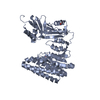
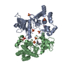
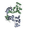
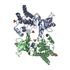
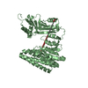
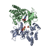
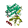
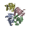
| Title | Crystal structure of the actin-crosslinking core of Arabidopsis fimbrin | ||||||
 Components Components | fimbrin-like protein | ||||||
 Keywords Keywords | STRUCTURAL PROTEIN / CALPONIN HOMOLOGY / F-ACTIN-BINDING DOMAIN (ABD) / F-ACTIN-CROSSLINKING / Structural Genomics / PSI / Protein Structure Initiative / New York SGX Research Center for Structural Genomics / NYSGXRC | ||||||
| Function / homology |  Function and homology information Function and homology informationactin filament network formation / actin filament bundle / actin filament bundle assembly / chloroplast / actin filament / circadian rhythm / actin filament binding / metal ion binding / cytoplasm Similarity search - Function | ||||||
| Biological species |  | ||||||
| Method |  X-RAY DIFFRACTION / SYNCHROTRON / MAD / Resolution: 2.4 Å X-RAY DIFFRACTION / SYNCHROTRON / MAD / Resolution: 2.4 Å | ||||||
 Authors Authors | Klein, M.G. / Shi, W. / Tseng, Y. / Wirtz, D. / Almo, S.C. / Burley, S.K. / New York SGX Research Center for Structural Genomics (NYSGXRC) | ||||||
 Citation Citation | Journal: Structure / Year: 2004 Title: Structure of the actin crosslinking core of fimbrin. Authors: Klein, M.G. / Shi, W. / Ramagopal, U. / Tseng, Y. / Wirtz, D. / Kovar, D.R. / Staiger, C.J. / Almo, S.C. | ||||||
| History |
|
- Structure visualization
| Structure viewer | Molecule: MolmilJmol/JSmol |
|---|
- Downloads & links
-Download
| PDBx/mmCIF format | 1pxy.cif.gz | 198.6 KB | Display | PDBx/mmCIF format |
|---|---|---|---|---|
| PDB format | pdb1pxy.ent.gz | 156.9 KB | Display | PDB format |
| PDBx/mmJSON format | 1pxy.json.gz | Tree view | PDBx/mmJSON format | |
| Others |  Other downloads Other downloads |
-Validation report
| Arichive directory | https://data.pdbj.org/pub/pdb/validation_reports/px/1pxyftp://data.pdbj.org/pub/pdb/validation_reports/px/1pxy | HTTPS FTP |
|---|
-Related structure data
| Related structure data |  1rt8C  1aoaS S: Starting model for refinement C: citing same article ( |
|---|---|
| Similar structure data | |
| Other databases |


-Links
 PDBj
PDBj- Assembly
| Deposited unit |
| ||||||||
|---|---|---|---|---|---|---|---|---|---|
| 1 |
| ||||||||
| 2 |
| ||||||||
| Unit cell |
|
-Components
| #1: Protein | Mass: 57547.645 Da / Num. of mol.: 2 / Fragment: actin-crosslinking core, residues 123-623 / Mutation: V240L Source method: isolated from a genetically manipulated source Source: (gene. exp.)  #2: Water | ChemComp-HOH / |  Mass: 18.015 Da / Num. of mol.: 190 / Source method: isolated from a natural source / Formula: H2O Mass: 18.015 Da / Num. of mol.: 190 / Source method: isolated from a natural source / Formula: H2O |
|---|
-Experimental details
-Experiment
| Experiment | Method: X-RAY DIFFRACTION / Number of used crystals: 2 |
|---|
- Sample preparation
| Crystal | Density Matthews: 2.9 Å3/Da / Density % sol: 57.53 % |
|---|---|
| Crystal grow | Temperature: 298 K / Method: vapor diffusion, hanging drop / pH: 8.2 Details: 6% PEG 8000, 100 mM Tris, 1mM DTT, pH 8.2, VAPOR DIFFUSION, HANGING DROP, temperature 298K |
-Data collection
| Diffraction | Mean temperature: 200 K | |||||||||||||||
|---|---|---|---|---|---|---|---|---|---|---|---|---|---|---|---|---|
| Diffraction source | Source: SYNCHROTRON / Site: NSLS  / Beamline: X9B / Wavelength: 0.984, 0.9786, 0.9789, 0.9560 / Beamline: X9B / Wavelength: 0.984, 0.9786, 0.9789, 0.9560 | |||||||||||||||
| Detector | Type: ADSC QUANTUM 4 / Detector: CCD / Date: Mar 1, 2001 | |||||||||||||||
| Radiation | Monochromator: Double flat crystal monochromator with fixed exit geometry Protocol: MAD / Monochromatic (M) / Laue (L): M / Scattering type: x-ray | |||||||||||||||
| Radiation wavelength |
| |||||||||||||||
| Reflection | Resolution: 2.4→30 Å / Num. all: 51046 / Num. obs: 44865 / % possible obs: 87.5 % / Observed criterion σ(F): 0 / Observed criterion σ(I): 0 / Redundancy: 5.97 % / Rsym value: 0.067 / Net I/σ(I): 20.71 | |||||||||||||||
| Reflection shell | Resolution: 2.4→2.49 Å / Redundancy: 4.1 % / Rmerge(I) obs: 0.315 / Mean I/σ(I) obs: 3.35 / Num. unique all: 4394 / Rsym value: 0.303 / % possible all: 86.7 |
- Processing
| Software |
| |||||||||||||||||||||||||
|---|---|---|---|---|---|---|---|---|---|---|---|---|---|---|---|---|---|---|---|---|---|---|---|---|---|---|
| Refinement | Method to determine structure: MAD Starting model: PDB ENTRY 1AOA Resolution: 2.4→20 Å / Cross valid method: THROUGHOUT / σ(F): 0 / Stereochemistry target values: Engh & Huber
| |||||||||||||||||||||||||
| Refinement step | Cycle: LAST / Resolution: 2.4→20 Å
| |||||||||||||||||||||||||
| LS refinement shell | Resolution: 2.4→2.49 Å
|
