Movie
Movie Controller


+ Open data

- Basic information
| Entry | Database: EMDB / ID: EMD-3708 | |||||||||
|---|---|---|---|---|---|---|---|---|---|---|
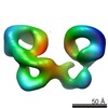
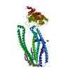
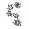
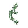
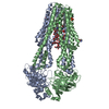
| Title | Negative-stain surface of SorCS1 monomer | |||||||||
 Map data Map data | Negative stain surface of mSorCS1 monomer | |||||||||
 Sample Sample |
| |||||||||
| Biological species |  | |||||||||
| Method | single particle reconstruction / negative staining / Resolution: 18.0 Å | |||||||||
 Authors Authors | Moeller A / Januliene D | |||||||||
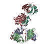
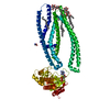
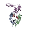
 Citation Citation | Journal: J Mol Biol / Year: 2017 Title: Hidden Twins: SorCS Neuroreceptors Form Stable Dimers. Authors: Dovile Januliene / Arulmani Manavalan / Peter Lund Ovesen / Karen-Marie Pedersen / Søren Thirup / Anders Nykjær / Arne Moeller /   Abstract: SorCS1, SorCS2 and SorCS3 belong to the Vps10p-domain family of multiligand receptors. Genetic and functional studies have linked SorCS receptors to psychiatric disorders, Alzheimer's disease and ...SorCS1, SorCS2 and SorCS3 belong to the Vps10p-domain family of multiligand receptors. Genetic and functional studies have linked SorCS receptors to psychiatric disorders, Alzheimer's disease and type 2 diabetes, demonstrating critical roles in neuronal functionality and metabolic control. Surprisingly, their structural composition has so far not been studied. Here we have characterized SorCS1, SorCS2 and SorCS3 using biochemical methods and electron microscopy. We found that their purified extracellular domains co-exist in stable dimeric and monomeric populations. This was supported by co-immunoprecipitation experiments, where membrane-bound dimers were successfully pulled down from cell lysate. While dimers were virtually unbreakable, dimerization of the monomeric population was promoted through enzymatic deglycosylation. We conclude that post-translational modifications, specifically the degree and pattern of glycosylation, regulate the oligomeric state of the protein. Hence, cells may dictate ligand specificity by controlling the ratio between monomers and dimers and, therefore, regulate the multiple functions of SorCS receptors. | |||||||||
| History |
|
- Structure visualization
| Movie |
 Movie viewer Movie viewer |
|---|---|
| Structure viewer | EM map: SurfViewMolmilJmol/JSmol |
| Supplemental images |
 UCSF Chimera
- Downloads & links
-EMDB archive
| Map data | emd_3708.map.gz | 80.7 KB | EMDB map data format | |
|---|---|---|---|---|
| Header (meta data) | emd-3708-v30.xmlemd-3708.xml | 8 KB 8 KB | Display Display | EMDB header |
| Images | 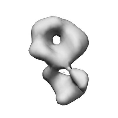 emd_3708.png emd_3708.png | 29.7 KB | ||
| Archive directory |  http://ftp.pdbj.org/pub/emdb/structures/EMD-3708ftp://ftp.pdbj.org/pub/emdb/structures/EMD-3708 http://ftp.pdbj.org/pub/emdb/structures/EMD-3708ftp://ftp.pdbj.org/pub/emdb/structures/EMD-3708 | HTTPS FTP |
-Related structure data


-Links
| EMDB pages | EMDB (EBI/PDBe) / EMDataResource |
|---|
-Map
| File | Download / File: emd_3708.map.gz / Format: CCP4 / Size: 85.9 KB / Type: IMAGE STORED AS FLOATING POINT NUMBER (4 BYTES) | ||||||||||||||||||||||||||||||||||||||||||||||||||||||||||||||||||||
|---|---|---|---|---|---|---|---|---|---|---|---|---|---|---|---|---|---|---|---|---|---|---|---|---|---|---|---|---|---|---|---|---|---|---|---|---|---|---|---|---|---|---|---|---|---|---|---|---|---|---|---|---|---|---|---|---|---|---|---|---|---|---|---|---|---|---|---|---|---|
| Annotation | Negative stain surface of mSorCS1 monomer | ||||||||||||||||||||||||||||||||||||||||||||||||||||||||||||||||||||
| Projections & slices | Image control
Images are generated by Spider. | ||||||||||||||||||||||||||||||||||||||||||||||||||||||||||||||||||||
| Voxel size | X=Y=Z: 6.29 Å | ||||||||||||||||||||||||||||||||||||||||||||||||||||||||||||||||||||
| Density |
| ||||||||||||||||||||||||||||||||||||||||||||||||||||||||||||||||||||
| Symmetry | Space group: 1 | ||||||||||||||||||||||||||||||||||||||||||||||||||||||||||||||||||||
| Details | EMDB XML:
CCP4 map header:
| ||||||||||||||||||||||||||||||||||||||||||||||||||||||||||||||||||||
 Z (Sec.)
 Y (Row.)
 X (Col.)


-Supplemental data
- Sample components
-Entire : SorCS1
| Entire | Name: SorCS1 |
|---|---|
| Components |
|
-Supramolecule #1: SorCS1
| Supramolecule | Name: SorCS1 / type: complex / ID: 1 / Parent: 0 |
|---|---|
| Source (natural) | Organism: |
| Recombinant expression | Organism:  Cricetulus griseus (Chinese hamster) Cricetulus griseus (Chinese hamster) |
| Molecular weight | Experimental: 140 KDa |
-Experimental details
-Structure determination
| Method | negative staining |
|---|---|
 Processing Processing | single particle reconstruction |
| Aggregation state | particle |
-Sample preparation
| Buffer | pH: 8 |
|---|---|
| Staining | Type: NEGATIVE / Material: Uranyl Formate |
- Electron microscopy
| Microscope | FEI TECNAI SPIRIT |
|---|---|
| Image recording | Film or detector model: TVIPS TEMCAM-F416 (4k x 4k) / Average electron dose: 8.0 e/Å2 |
| Electron beam | Acceleration voltage: 120 kV / Electron source: LAB6 |
| Electron optics | Illumination mode: FLOOD BEAM / Imaging mode: BRIGHT FIELD |
| Experimental equipment |  Model: Tecnai Spirit / Image courtesy: FEI Company |
-Image processing
| Startup model | Type of model: PDB ENTRY PDB model - PDB ID: |
|---|---|
| Final reconstruction | Applied symmetry - Point group: C1 (asymmetric) / Resolution.type: BY AUTHOR / Resolution: 18.0 Å / Resolution method: FSC 0.143 CUT-OFF / Number images used: 34000 |
| Initial angle assignment | Type: PROJECTION MATCHING |
| Final angle assignment | Type: PROJECTION MATCHING |