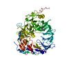
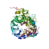
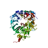
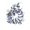
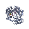
Mass: 76774.703 Da / Num. of mol.: 1 / Fragment: UNP residues 78-756 Source method: isolated from a genetically manipulated source Source: (gene. exp.) Homo sapiens (human) / Gene: SORT1 / Production host: Cricetulus griseus (Chinese hamster) / Strain (production host): CHO-K1 / References: UniProt: Q99523
#2: Protein/peptide
Neurotensin
Mass: 1675.948 Da / Num. of mol.: 1 / Fragment: UNP residues 151-163 / Source method: obtained synthetically Details: Synthetic peptide purchased from Sigma. It represents the naturally occurring form of neurotensin in human, residues 151-163 of UniProt entry P30990, NEUT_HUMAN References: UniProt: P30990
Mass: 18.015 Da / Num. of mol.: 305 / Source method: isolated from a natural source / Formula: H2O
-
Details
Compound details
AUTHORS STATE THAT THE GLYCOSYLATIONS ARE COVALENTLY ATTACHED TO THE ASPARAGINES, THEREFORE THEY ...AUTHORS STATE THAT THE GLYCOSYLATIONS ARE COVALENTLY ATTACHED TO THE ASPARAGINES, THEREFORE THEY SHOULD NOT BE DESCRIBED AS SITE RECORDS. INSTEAD, WHAT SHOULD BE SHOWN IN SITE RECORDS, IS THE NEUROTENSIN BINDING SITE.
Has protein modification
Y
Sequence details
IN VIVO, NEUROTENSIN (ENTITY 2) IS PRODUCED BY PROTEOLYTIC CLEAVAGE OF THE GENE PRODUCT, AND THE ...IN VIVO, NEUROTENSIN (ENTITY 2) IS PRODUCED BY PROTEOLYTIC CLEAVAGE OF THE GENE PRODUCT, AND THE RESULTING N-TERMINAL GLUTAMATE IS NATURALLY CONVERTED TO PYROGLUTAMATE.
-
Experimental details
-
Experiment
Experiment
Method: X-RAY DIFFRACTION / Number of used crystals: 1
-
Sample preparation
Crystal
Density Matthews: 2.79 Å3/Da / Density % sol: 55.96 %
Crystal grow
Temperature: 293 K / Method: vapor diffusion, sitting drop / pH: 7.8 Details: 19% PEG 6000, 600 mM NaCl, 3% Glycerol, 100 mM HEPES, 93 mM Tris-HCl, pH 7.8, VAPOR DIFFUSION, SITTING DROP, temperature 293K
In the structure databanks used in Yorodumi, some data are registered as the other names, "COVID-19 virus" and "2019-nCoV". Here are the details of the virus and the list of structure data.
Jan 31, 2019. EMDB accession codes are about to change! (news from PDBe EMDB page)
EMDB accession codes are about to change! (news from PDBe EMDB page)
The allocation of 4 digits for EMDB accession codes will soon come to an end. Whilst these codes will remain in use, new EMDB accession codes will include an additional digit and will expand incrementally as the available range of codes is exhausted. The current 4-digit format prefixed with “EMD-” (i.e. EMD-XXXX) will advance to a 5-digit format (i.e. EMD-XXXXX), and so on. It is currently estimated that the 4-digit codes will be depleted around Spring 2019, at which point the 5-digit format will come into force.
The EM Navigator/Yorodumi systems omit the EMD- prefix.
Related info.:Q: What is EMD? / ID/Accession-code notation in Yorodumi/EM Navigator
Yorodumi is a browser for structure data from EMDB, PDB, SASBDB, etc.
This page is also the successor to EM Navigator detail page, and also detail information page/front-end page for Omokage search.
The word "yorodu" (or yorozu) is an old Japanese word meaning "ten thousand". "mi" (miru) is to see.
Related info.:EMDB / PDB / SASBDB / Comparison of 3 databanks / Yorodumi Search / Aug 31, 2016. New EM Navigator & Yorodumi / Yorodumi Papers / Jmol/JSmol / Function and homology information / Changes in new EM Navigator and Yorodumi
 Movie
Movie Controller

 Yorodumi
 Open data
 Basic information
 Components
 Keywords
 Function and homology information
 Homo sapiens (human)
 X-RAY DIFFRACTION /
  Authors
 Citation
 Structure visualization
 Downloads & links
 Other downloads
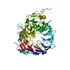


 PDBj
PDBj


 Assembly

 Cricetulus griseus (Chinese hamster) / Strain (production host): CHO-K1 / References: UniProt: Q99523


 Mass: 150.173 Da / Num. of mol.: 1 / Source method: obtained synthetically / Formula: C6H14O4
 Mass: 92.094 Da / Num. of mol.: 1 / Source method: obtained synthetically / Formula: C3H8O3
 Sample preparation
 / Beamline: X06SA / Wavelength: 0.95008 Å
 Processing