 Movie
Movie Controller


+ Open data

- Basic information
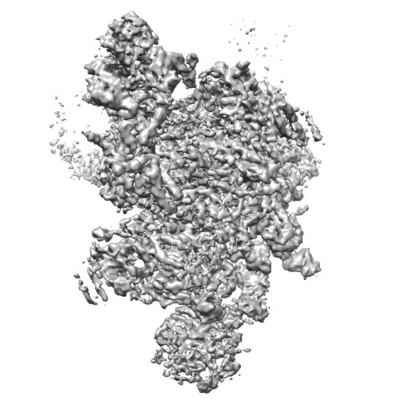
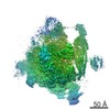
| Entry | Database: EMDB / ID: EMD-3541 | ||||||||||||
|---|---|---|---|---|---|---|---|---|---|---|---|---|---|
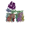
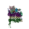
| Title | Structure of a spliceosome remodeled for exon ligation | ||||||||||||
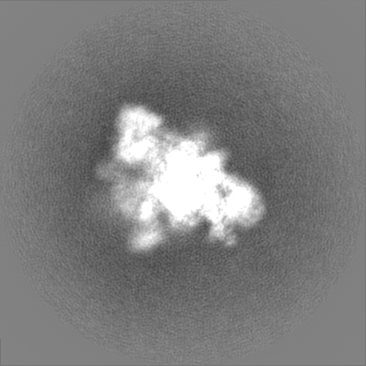
 Map data Map data | Map of the spliceosomal C-star complex resulting from focussed classification on Prp22 | ||||||||||||
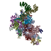
 Sample Sample |
| ||||||||||||
 Keywords Keywords | pre-mRNA splicing / trans-esterification / lariat intermediate / complex C-star / Splicing | ||||||||||||
| Function / homology |  Function and homology information Function and homology informationU2-type post-spliceosomal complex / mRNA branch site recognition / spliceosomal complex disassembly / U2-type post-mRNA release spliceosomal complex / cellular bud site selection / pre-mRNA 3'-splice site binding / post-mRNA release spliceosomal complex / generation of catalytic spliceosome for first transesterification step / cis assembly of pre-catalytic spliceosome / nuclear mRNA surveillance ...U2-type post-spliceosomal complex / mRNA branch site recognition / spliceosomal complex disassembly / U2-type post-mRNA release spliceosomal complex / cellular bud site selection / pre-mRNA 3'-splice site binding / post-mRNA release spliceosomal complex / generation of catalytic spliceosome for first transesterification step / cis assembly of pre-catalytic spliceosome / nuclear mRNA surveillance / spliceosome conformational change to release U4 (or U4atac) and U1 (or U11) / U4/U6 snRNP / 7-methylguanosine cap hypermethylation / pre-mRNA binding / U2-type catalytic step 1 spliceosome / pICln-Sm protein complex / small nuclear ribonucleoprotein complex / SMN-Sm protein complex / spliceosomal tri-snRNP complex / splicing factor binding / snRNP binding / commitment complex / mRNA cis splicing, via spliceosome / U2-type prespliceosome assembly / U2-type catalytic step 2 spliceosome / U2-type spliceosomal complex / U1 snRNP / U2 snRNP / U4 snRNP / U2-type prespliceosome / poly(U) RNA binding / generation of catalytic spliceosome for second transesterification step / precatalytic spliceosome / mRNA 5'-splice site recognition / Formation of TC-NER Pre-Incision Complex / mRNA 3'-splice site recognition / spliceosomal complex assembly / Gap-filling DNA repair synthesis and ligation in TC-NER / spliceosomal tri-snRNP complex assembly / Prp19 complex / DNA replication origin binding / U5 snRNP / Dual incision in TC-NER / U5 snRNA binding / pre-mRNA intronic binding / DNA replication initiation / spliceosomal snRNP assembly / U2 snRNA binding / U6 snRNA binding / protein K63-linked ubiquitination / U1 snRNA binding / U4/U6 x U5 tri-snRNP complex / catalytic step 2 spliceosome / positive regulation of cell cycle / nuclear periphery / positive regulation of RNA splicing / spliceosomal complex / mRNA splicing, via spliceosome / RING-type E3 ubiquitin transferase / metallopeptidase activity / ubiquitin-protein transferase activity / ubiquitin protein ligase activity / RNA helicase activity / RNA helicase / DNA repair / mRNA binding / GTPase activity / chromatin binding / chromatin / GTP binding / ATP hydrolysis activity / mitochondrion / DNA binding / RNA binding / zinc ion binding / ATP binding / identical protein binding / nucleus / cytoplasm / cytosol Similarity search - Function | ||||||||||||
| Biological species |  | ||||||||||||
| Method | single particle reconstruction / cryo EM / Resolution: 4.17 Å | ||||||||||||
 Authors Authors | Fica SM / Oubridge C | ||||||||||||
| Funding support |  United Kingdom, 3 items United Kingdom, 3 items
| ||||||||||||
 Citation Citation | Journal: Nature / Year: 2016 Title: Cryo-EM structure of the spliceosome immediately after branching. Authors: Wojciech P Galej / Max E Wilkinson / Sebastian M Fica / Chris Oubridge / Andrew J Newman / Kiyoshi Nagai / Abstract: Precursor mRNA (pre-mRNA) splicing proceeds by two consecutive transesterification reactions via a lariat-intron intermediate. Here we present the 3.8 Å cryo-electron microscopy structure of the ...Precursor mRNA (pre-mRNA) splicing proceeds by two consecutive transesterification reactions via a lariat-intron intermediate. Here we present the 3.8 Å cryo-electron microscopy structure of the spliceosome immediately after lariat formation. The 5'-splice site is cleaved but remains close to the catalytic Mg site in the U2/U6 small nuclear RNA (snRNA) triplex, and the 5'-phosphate of the intron nucleotide G(+1) is linked to the branch adenosine 2'OH. The 5'-exon is held between the Prp8 amino-terminal and linker domains, and base-pairs with U5 snRNA loop 1. Non-Watson-Crick interactions between the branch helix and 5'-splice site dock the branch adenosine into the active site, while intron nucleotides +3 to +6 base-pair with the U6 snRNA ACAGAGA sequence. Isy1 and the step-one factors Yju2 and Cwc25 stabilize docking of the branch helix. The intron downstream of the branch site emerges between the Prp8 reverse transcriptase and linker domains and extends towards the Prp16 helicase, suggesting a plausible mechanism of remodelling before exon ligation. | ||||||||||||
| History |
|
- Structure visualization
| Movie |
Movie viewer |
|---|---|
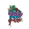
| Structure viewer | EM map: SurfViewMolmilJmol/JSmol |
| Supplemental images |
- Downloads & links
-EMDB archive
| Map data | emd_3541.map.gz | 248.2 MB | EMDB map data format | |
|---|---|---|---|---|
| Header (meta data) | emd-3541-v30.xmlemd-3541.xml | 78.3 KB 78.3 KB | Display Display | EMDB header |
| FSC (resolution estimation) | emd_3541_fsc_1.xmlemd_3541_fsc_2.xml | 14.5 KB 14.5 KB | Display Display | FSC data file |
| Images |  emd_3541_1.png emd_3541_1.png emd_3541_2.png emd_3541_2.png | 63.7 KB 62.6 KB | ||
| Filedesc metadata | emd-3541.cif.gz | 20.2 KB | ||
| Others | emd_3541_additional.map.gz | 243.9 MB | ||
| Archive directory |  http://ftp.pdbj.org/pub/emdb/structures/EMD-3541ftp://ftp.pdbj.org/pub/emdb/structures/EMD-3541 http://ftp.pdbj.org/pub/emdb/structures/EMD-3541ftp://ftp.pdbj.org/pub/emdb/structures/EMD-3541 | HTTPS FTP |
-Related structure data
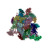
| Related structure data |  5mq0MC  3539C 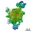 3542C 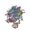 5mpsC M: atomic model generated by this map C: citing same article ( |
|---|---|
| Similar structure data | |
| EM raw data | EMPIAR-10687 (Title: Yeast C, Ci, C*, and P complex spliceosomes / Data size: 8.9 TB Data #1: Unaligned movies of C-complex spliceosome with 3' splice site AG to AC mutation (Dataset 1) [micrographs - multiframe] Data #2: Unaligned movies of C and C*-complex spliceosomes with 3' splice site AG to AdG mutation (Dataset 2) [micrographs - multiframe] Data #3: Unaligned movies of C and C*-complex spliceosomes with 3' splice site AG to AdG mutation (Dataset 3) [micrographs - multiframe] Data #4: Aligned movies of C-complex spliceosomes with cold-sensitive prp16-302 mutation, purified with Cwc25 (Dataset 4) [micrographs - multiframe] Data #5: Unaligned movies of C-complex spliceosomes with cold-sensitive prp16-302 mutation, purified with Cwc25 and incubated with ATP and Mg (Dataset 5) [micrographs - multiframe] Data #6: Unaligned movies of C, C*, and P-complex spliceosomes with dominant-negative Prp22 mutation K512A, purified with Slu7 (Dataset 6) [micrographs - multiframe] Data #7: Unaligned movies of P-complex spliceosomes with dominant-negative Prp22 mutation K512A, treated with anti-3'exon RNaseH oligo, purified in presence of Mg (Dataset 9) [micrographs - single frame] Data #8: Selected C-complex particles after polishing [picked particles - single frame - processed] Data #9: Selected P-complex particles after polishing [picked particles - single frame - processed] Data #10: Various signal subtractions for C- and P-complex spliceosomes [picked particles - single frame - processed]) |
-Links
| EMDB pages | EMDB (EBI/PDBe) / EMDataResource |
|---|---|
| Related items in Molecule of the Month |


-Map
| File | Download / File: emd_3541.map.gz / Format: CCP4 / Size: 266.8 MB / Type: IMAGE STORED AS FLOATING POINT NUMBER (4 BYTES) | ||||||||||||||||||||||||||||||||||||||||||||||||||||||||||||
|---|---|---|---|---|---|---|---|---|---|---|---|---|---|---|---|---|---|---|---|---|---|---|---|---|---|---|---|---|---|---|---|---|---|---|---|---|---|---|---|---|---|---|---|---|---|---|---|---|---|---|---|---|---|---|---|---|---|---|---|---|---|
| Annotation | Map of the spliceosomal C-star complex resulting from focussed classification on Prp22 | ||||||||||||||||||||||||||||||||||||||||||||||||||||||||||||
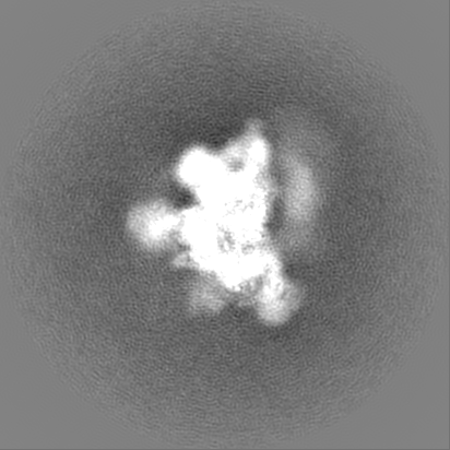
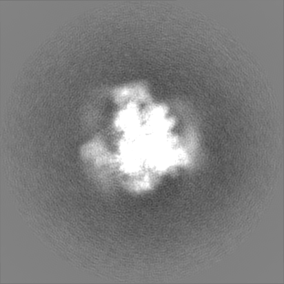
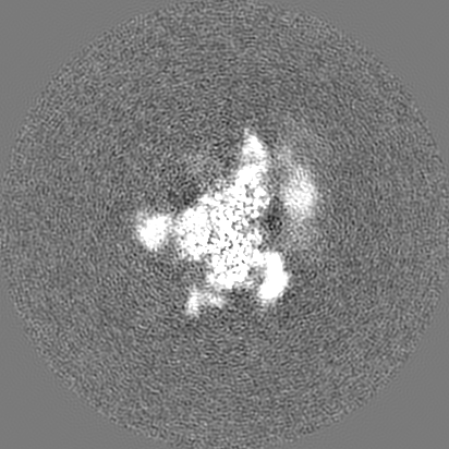
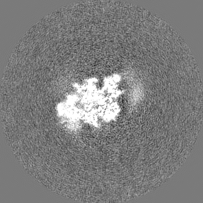
| Projections & slices | Image control
Images are generated by Spider. | ||||||||||||||||||||||||||||||||||||||||||||||||||||||||||||
| Voxel size | X=Y=Z: 1.43 Å | ||||||||||||||||||||||||||||||||||||||||||||||||||||||||||||
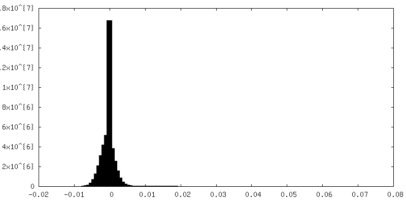
| Density |
| ||||||||||||||||||||||||||||||||||||||||||||||||||||||||||||
| Symmetry | Space group: 1 | ||||||||||||||||||||||||||||||||||||||||||||||||||||||||||||
| Details | EMDB XML:
CCP4 map header:
| ||||||||||||||||||||||||||||||||||||||||||||||||||||||||||||
 Z (Sec.)
 Y (Row.)
 X (Col.)


-Supplemental data
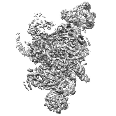
-Additional map: Map of the spliceosomal C-star complex resulting from...
| File | emd_3541_additional.map | ||||||||||||
|---|---|---|---|---|---|---|---|---|---|---|---|---|---|
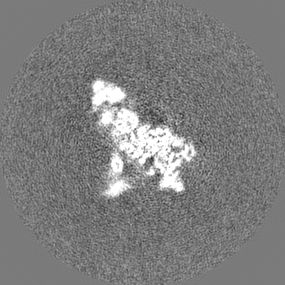
| Annotation | Map of the spliceosomal C-star complex resulting from focussed classification on peripheral regions of U2 snRNP | ||||||||||||
| Projections & Slices |
| ||||||||||||
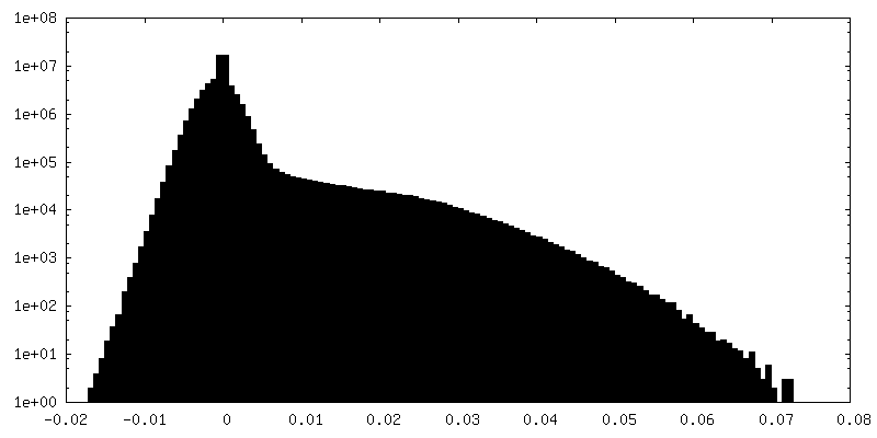
| Density Histograms |
- Sample components
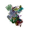
+Entire : Saccharomyces cerevisiae spliceosome. Complex C just after Prp16-...
+Supramolecule #1: Saccharomyces cerevisiae spliceosome. Complex C just after Prp16-...
+Macromolecule #1: Yeast UBC4 gene for ubiquitin-conjugating enzyme
+Macromolecule #2: 5'-EXON OF UBC4 PRE-MRNA
+Macromolecule #3: S.cerevisiae chromosome II reading frame ORF YBR230c
+Macromolecule #4: Saccharomyces cerevisiae strain T.52_2H chromosome XII sequence
+Macromolecule #5: Saccharomyces cerevisiae strain WI_C_MBSP_4 chromosome VII sequence
+Macromolecule #34: 3'-EXON OF UBC4 PRE-MRNA, BOUND BY PRP22 HELICASE
+Macromolecule #6: Pre-mRNA-splicing factor 8
+Macromolecule #7: Pre-mRNA-splicing factor SNU114
+Macromolecule #8: Pre-mRNA-splicing factor CWC22
+Macromolecule #9: Pre-mRNA-splicing factor PRP46
+Macromolecule #10: Pre-mRNA-processing protein 45
+Macromolecule #11: Pre-mRNA-splicing factor BUD31
+Macromolecule #12: Pre-mRNA-splicing factor CWC2
+Macromolecule #13: Pre-mRNA-splicing factor SLT11
+Macromolecule #14: Pre-mRNA-splicing factor CEF1
+Macromolecule #15: Pre-mRNA-splicing factor CWC15
+Macromolecule #16: Pre-mRNA-splicing factor CWC21
+Macromolecule #17: Pre-mRNA-splicing factor CLF1
+Macromolecule #18: Pre-mRNA-splicing factor SYF1,PRE-MRNA-SPLICING FACTOR SYF1
+Macromolecule #19: Pre-mRNA-splicing factor 18
+Macromolecule #20: Pre-mRNA-splicing factor SLU7
+Macromolecule #21: Pre-mRNA-processing factor 17
+Macromolecule #22: UNKNOWN PROTEIN
+Macromolecule #23: Pre-mRNA-splicing factor SYF2
+Macromolecule #24: Small nuclear ribonucleoprotein-associated protein B
+Macromolecule #25: Small nuclear ribonucleoprotein Sm D3
+Macromolecule #26: Small nuclear ribonucleoprotein E
+Macromolecule #27: Small nuclear ribonucleoprotein F
+Macromolecule #28: Small nuclear ribonucleoprotein G
+Macromolecule #29: Small nuclear ribonucleoprotein Sm D1
+Macromolecule #30: Small nuclear ribonucleoprotein Sm D2
+Macromolecule #31: Pre-mRNA-splicing factor ATP-dependent RNA helicase PRP22
+Macromolecule #32: U2 small nuclear ribonucleoprotein A'
+Macromolecule #33: U2 small nuclear ribonucleoprotein B''
+Macromolecule #35: Pre-mRNA-splicing factor SNT309
+Macromolecule #36: Pre-mRNA-processing factor 19
+Macromolecule #37: MAGNESIUM ION
+Macromolecule #38: POTASSIUM ION
+Macromolecule #39: INOSITOL HEXAKISPHOSPHATE

+Macromolecule #40: GUANOSINE-5'-TRIPHOSPHATE

+Macromolecule #41: ZINC ION
-Experimental details
-Structure determination
| Method | cryo EM |
|---|---|
 Processing Processing | single particle reconstruction |
| Aggregation state | particle |
-Sample preparation
| Concentration | 0.3 mg/mL | ||||||||||||||||||
|---|---|---|---|---|---|---|---|---|---|---|---|---|---|---|---|---|---|---|---|
| Buffer | pH: 7.9 Component:
Details: NP-40 is also called IGEPAL CA-630 | ||||||||||||||||||
| Grid | Model: Quantifoil R1.2/1.3 / Material: COPPER / Mesh: 400 / Support film - Material: CARBON / Support film - topology: HOLEY ARRAY / Support film - Film thickness: 6 / Pretreatment - Type: GLOW DISCHARGE / Pretreatment - Time: 30 sec. / Pretreatment - Atmosphere: AIR / Pretreatment - Pressure: 20.0 kPa | ||||||||||||||||||
| Vitrification | Cryogen name: ETHANE / Chamber humidity: 100 % / Chamber temperature: 277 K / Instrument: FEI VITROBOT MARK III Details: 3.5 microlitres sample were applied to the grid, left for 25 seconds and then blotted for 3.0-3.5 seconds before plunging.. |
- Electron microscopy
| Microscope | FEI TITAN KRIOS |
|---|---|
| Specialist optics | Energy filter - Name: GIF Quantum |
| Details | GIF Quantum energy filter, 20 eV slit width |
| Image recording | Film or detector model: GATAN K2 SUMMIT (4k x 4k) / Detector mode: SUPER-RESOLUTION / Number real images: 3596 / Average exposure time: 0.8 sec. / Average electron dose: 2.0 e/Å2 Details: Total dose: 40 electrons/Angstrom^2 over 16 seconds. 20 movie frames collected at 1.25 frames per second. |
| Electron beam | Acceleration voltage: 300 kV / Electron source:  FIELD EMISSION GUN FIELD EMISSION GUN |
| Electron optics | Illumination mode: FLOOD BEAM / Imaging mode: BRIGHT FIELD / Nominal defocus max: 4.5 µm / Nominal defocus min: 0.5 µm / Nominal magnification: 81000 |
| Sample stage | Specimen holder model: FEI TITAN KRIOS AUTOGRID HOLDER / Cooling holder cryogen: NITROGEN |
| Experimental equipment |  Model: Titan Krios / Image courtesy: FEI Company |
+Image processing

-Atomic model buiding 1
| Details | Used secondary structure restraints generated in ProSMART and LibG. |
|---|---|
| Refinement | Space: RECIPROCAL / Protocol: FLEXIBLE FIT / Overall B value: 330 / Target criteria: Fourier Shell Correlation |
| Output model | PDB-5mq0: |
