Movie
Movie Controller
 Structure viewers
 About Yorodumi Papers


+Search query
-Structure paper
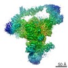
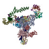
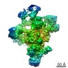
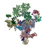
| Title | Cryo-EM structure of the spliceosome immediately after branching. |
|---|---|
| Journal, issue, pages | Nature, Vol. 537, Issue 7619, Page 197-201, Year 2016 |
| Publish date | Sep 8, 2016 |
 Authors Authors | Wojciech P Galej / Max E Wilkinson / Sebastian M Fica / Chris Oubridge / Andrew J Newman / Kiyoshi Nagai /  |
| PubMed Abstract | Precursor mRNA (pre-mRNA) splicing proceeds by two consecutive transesterification reactions via a lariat-intron intermediate. Here we present the 3.8 Å cryo-electron microscopy structure of the ...Precursor mRNA (pre-mRNA) splicing proceeds by two consecutive transesterification reactions via a lariat-intron intermediate. Here we present the 3.8 Å cryo-electron microscopy structure of the spliceosome immediately after lariat formation. The 5'-splice site is cleaved but remains close to the catalytic Mg site in the U2/U6 small nuclear RNA (snRNA) triplex, and the 5'-phosphate of the intron nucleotide G(+1) is linked to the branch adenosine 2'OH. The 5'-exon is held between the Prp8 amino-terminal and linker domains, and base-pairs with U5 snRNA loop 1. Non-Watson-Crick interactions between the branch helix and 5'-splice site dock the branch adenosine into the active site, while intron nucleotides +3 to +6 base-pair with the U6 snRNA ACAGAGA sequence. Isy1 and the step-one factors Yju2 and Cwc25 stabilize docking of the branch helix. The intron downstream of the branch site emerges between the Prp8 reverse transcriptase and linker domains and extends towards the Prp16 helicase, suggesting a plausible mechanism of remodelling before exon ligation. |
 External links External links | Nature / PubMed:27459055 / PubMed Central |
| Methods | EM (single particle) |
| Resolution | 3.8 - 10.0 Å |
| Structure data | EMDB-4055: Overall map of the yeast spliceosome immediately after branching 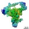 EMDB-4056: EMDB-4057: Structure of the yeast spliceosome immediately after branching. 3D class containing helicase module. 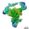 EMDB-4058: 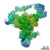 EMDB-4059: |
| Chemicals |  ChemComp-MG:  ChemComp-ZN:  ChemComp-GTP: |
| Source |
|
 Keywords Keywords | SPLICING / spliceosome / snRNP / pre-mRNA splicing / trans-esterification / lariat intermediate / complex C |