 ムービー
ムービー コントローラー


+ データを開く

- 基本情報
基本情報
| 登録情報 | データベース: EMDB / ID: EMD-31422 | |||||||||
|---|---|---|---|---|---|---|---|---|---|---|
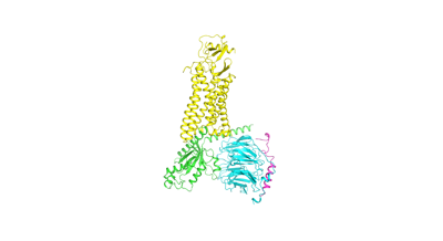
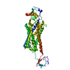
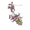
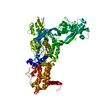
| タイトル | Cryo-EM structure of the chemokine receptor CCR5 in complex with MIP-1a and Gi | |||||||||
 マップデータ マップデータ | ||||||||||
 試料 試料 |
| |||||||||
| 機能・相同性 |  機能・相同性情報 機能・相同性情報chemokine (C-C motif) ligand 5 binding / granulocyte chemotaxis / CCR1 chemokine receptor binding / positive regulation of microglial cell migration / positive regulation of natural killer cell chemotaxis / negative regulation of macrophage apoptotic process / signaling / regulation of behavior / chemokine receptor activity / astrocyte cell migration ...chemokine (C-C motif) ligand 5 binding / granulocyte chemotaxis / CCR1 chemokine receptor binding / positive regulation of microglial cell migration / positive regulation of natural killer cell chemotaxis / negative regulation of macrophage apoptotic process / signaling / regulation of behavior / chemokine receptor activity / astrocyte cell migration / CCR5 chemokine receptor binding / eosinophil degranulation / regulation of sensory perception of pain / negative regulation of bone mineralization / CCR chemokine receptor binding / positive regulation of microglial cell activation / lymphocyte chemotaxis / cell activation / phosphatidylinositol phospholipase C activity / C-C chemokine receptor activity / T cell chemotaxis / C-C chemokine binding / positive regulation of calcium ion transport / eosinophil chemotaxis / response to cholesterol / chemokine-mediated signaling pathway / Chemokine receptors bind chemokines / release of sequestered calcium ion into cytosol by sarcoplasmic reticulum / chemokine activity / dendritic cell chemotaxis / phospholipase activator activity / positive regulation of calcium ion import / exocytosis / chemoattractant activity / negative regulation of osteoclast differentiation / macrophage chemotaxis / Interleukin-10 signaling / monocyte chemotaxis / negative regulation by host of viral transcription / Adenylate cyclase inhibitory pathway / positive regulation of protein localization to cell cortex / Binding and entry of HIV virion / cellular response to interleukin-1 / regulation of cAMP-mediated signaling / cellular defense response / D2 dopamine receptor binding / G protein-coupled serotonin receptor binding / coreceptor activity / positive regulation of calcium-mediated signaling / regulation of mitotic spindle organization / cellular response to forskolin / cytoskeleton organization / adenylate cyclase-inhibiting G protein-coupled receptor signaling pathway / neutrophil chemotaxis / cell chemotaxis / positive regulation of interleukin-1 beta production / Regulation of insulin secretion / G protein-coupled receptor binding / calcium-mediated signaling / G-protein beta/gamma-subunit complex binding / Olfactory Signaling Pathway / Activation of the phototransduction cascade / adenylate cyclase-modulating G protein-coupled receptor signaling pathway / G beta:gamma signalling through PLC beta / Presynaptic function of Kainate receptors / Thromboxane signalling through TP receptor / G-protein activation / G protein-coupled acetylcholine receptor signaling pathway / Activation of G protein gated Potassium channels / Inhibition of voltage gated Ca2+ channels via Gbeta/gamma subunits / Prostacyclin signalling through prostacyclin receptor / Glucagon signaling in metabolic regulation / G beta:gamma signalling through CDC42 / response to toxic substance / ADP signalling through P2Y purinoceptor 12 / G beta:gamma signalling through BTK / response to peptide hormone / Synthesis, secretion, and inactivation of Glucagon-like Peptide-1 (GLP-1) / Sensory perception of sweet, bitter, and umami (glutamate) taste / photoreceptor disc membrane / Adrenaline,noradrenaline inhibits insulin secretion / Glucagon-type ligand receptors / cellular response to type II interferon / Vasopressin regulates renal water homeostasis via Aquaporins / intracellular calcium ion homeostasis / G alpha (z) signalling events / positive regulation of inflammatory response / cellular response to catecholamine stimulus / Glucagon-like Peptide-1 (GLP1) regulates insulin secretion / ADORA2B mediated anti-inflammatory cytokines production / osteoblast differentiation / sensory perception of taste / ADP signalling through P2Y purinoceptor 1 / adenylate cyclase-activating dopamine receptor signaling pathway / G beta:gamma signalling through PI3Kgamma / cellular response to prostaglandin E stimulus / Cooperation of PDCL (PhLP1) and TRiC/CCT in G-protein beta folding / calcium ion transport / GPER1 signaling / GDP binding 類似検索 - 分子機能 | |||||||||
| 生物種 |  Homo sapiens (ヒト) Homo sapiens (ヒト) | |||||||||
| 手法 | 単粒子再構成法 / クライオ電子顕微鏡法 / 解像度: 2.9 Å | |||||||||
 データ登録者 データ登録者 | Zhang H / Chen K / Tan Q / Han S / Zhu Y / Zhao Q / Wu B | |||||||||
| 資金援助 |  中国, 2件 中国, 2件
| |||||||||
 引用 引用 | ジャーナル: Nat Commun / 年: 2021 タイトル: Structural basis for chemokine recognition and receptor activation of chemokine receptor CCR5. 著者: Hui Zhang / Kun Chen / Qiuxiang Tan / Qiang Shao / Shuo Han / Chenhui Zhang / Cuiying Yi / Xiaojing Chu / Ya Zhu / Yechun Xu / Qiang Zhao / Beili Wu / 要旨: The chemokine receptor CCR5 plays a vital role in immune surveillance and inflammation. However, molecular details that govern its endogenous chemokine recognition and receptor activation remain ...The chemokine receptor CCR5 plays a vital role in immune surveillance and inflammation. However, molecular details that govern its endogenous chemokine recognition and receptor activation remain elusive. Here we report three cryo-electron microscopy structures of G protein-coupled CCR5 in a ligand-free state and in complex with the chemokine MIP-1α or RANTES, as well as the crystal structure of MIP-1α-bound CCR5. These structures reveal distinct binding modes of the two chemokines and a specific accommodate pattern of the chemokine for the distal N terminus of CCR5. Together with functional data, the structures demonstrate that chemokine-induced rearrangement of toggle switch and plasticity of the receptor extracellular region are critical for receptor activation, while a conserved tryptophan residue in helix II acts as a trigger of receptor constitutive activation. | |||||||||
| 履歴 |
|
- 構造の表示
構造の表示
| ムービー |
ムービービューア |
|---|---|
| 構造ビューア | EMマップ: SurfViewMolmilJmol/JSmol |
| 添付画像 |
- ダウンロードとリンク
-EMDBアーカイブ
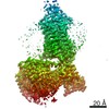
| マップデータ | emd_31422.map.gz | 59.8 MB | EMDBマップデータ形式 | |
|---|---|---|---|---|
| ヘッダ (付随情報) | emd-31422-v30.xmlemd-31422.xml | 13.7 KB 13.7 KB | 表示 表示 | EMDBヘッダ |
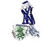
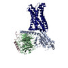
| 画像 |  emd_31422.png emd_31422.png | 26 KB | ||
| アーカイブディレクトリ |  http://ftp.pdbj.org/pub/emdb/structures/EMD-31422ftp://ftp.pdbj.org/pub/emdb/structures/EMD-31422 http://ftp.pdbj.org/pub/emdb/structures/EMD-31422ftp://ftp.pdbj.org/pub/emdb/structures/EMD-31422 | HTTPS FTP |
-検証レポート
| 文書・要旨 | emd_31422_validation.pdf.gz | 471.7 KB | 表示 | EMDB検証レポート |
|---|---|---|---|---|
| 文書・詳細版 | emd_31422_full_validation.pdf.gz | 471.3 KB | 表示 | |
| XML形式データ | emd_31422_validation.xml.gz | 6 KB | 表示 | |
| CIF形式データ | emd_31422_validation.cif.gz | 6.8 KB | 表示 | |
| アーカイブディレクトリ | https://ftp.pdbj.org/pub/emdb/validation_reports/EMD-31422ftp://ftp.pdbj.org/pub/emdb/validation_reports/EMD-31422 | HTTPS FTP |
-関連構造データ


-リンク
| EMDBのページ | EMDB (EBI/PDBe) / EMDataResource |
|---|---|
| 「今月の分子」の関連する項目 |


-マップ
| ファイル | ダウンロード / ファイル: emd_31422.map.gz / 形式: CCP4 / 大きさ: 64 MB / タイプ: IMAGE STORED AS FLOATING POINT NUMBER (4 BYTES) | ||||||||||||||||||||||||||||||||||||||||||||||||||||||||||||||||||||
|---|---|---|---|---|---|---|---|---|---|---|---|---|---|---|---|---|---|---|---|---|---|---|---|---|---|---|---|---|---|---|---|---|---|---|---|---|---|---|---|---|---|---|---|---|---|---|---|---|---|---|---|---|---|---|---|---|---|---|---|---|---|---|---|---|---|---|---|---|---|
| ボクセルのサイズ | X=Y=Z: 1.045 Å | ||||||||||||||||||||||||||||||||||||||||||||||||||||||||||||||||||||
| 密度 |
| ||||||||||||||||||||||||||||||||||||||||||||||||||||||||||||||||||||
| 対称性 | 空間群: 1 | ||||||||||||||||||||||||||||||||||||||||||||||||||||||||||||||||||||
| 詳細 | EMDB XML:
CCP4マップ ヘッダ情報:
| ||||||||||||||||||||||||||||||||||||||||||||||||||||||||||||||||||||
-添付データ
- 試料の構成要素
-全体 : Chemokine receptor CCR5 in complex with MIP-1a and Gi
| 全体 | 名称: Chemokine receptor CCR5 in complex with MIP-1a and Gi |
|---|---|
| 要素 |
|
-超分子 #1: Chemokine receptor CCR5 in complex with MIP-1a and Gi
| 超分子 | 名称: Chemokine receptor CCR5 in complex with MIP-1a and Gi タイプ: complex / ID: 1 / 親要素: 0 / 含まれる分子: all |
|---|---|
| 由来(天然) | 生物種: Homo sapiens (ヒト) |
| 組換発現 | 生物種:   Spodoptera frugiperda (ツマジロクサヨトウ) Spodoptera frugiperda (ツマジロクサヨトウ) |
-分子 #1: C-C motif chemokine 3,C-C chemokine receptor type 5
| 分子 | 名称: C-C motif chemokine 3,C-C chemokine receptor type 5 / タイプ: protein_or_peptide / ID: 1 / 詳細: Fusion protein of MIP-1a and CCR5 / コピー数: 1 / 光学異性体: LEVO |
|---|---|
| 由来(天然) | 生物種: Homo sapiens (ヒト) |
| 分子量 | 理論値: 51.241047 KDa |
| 組換発現 | 生物種: Spodoptera frugiperda (ツマジロクサヨトウ) |
| 配列 | 文字列: SLAADTPTAC CFSYCSRQIP QNFIADYFET SSQCSKPGVI FLTKRSRQVC ADPSEEWVQK YVSDLELSAG SGSGSGSGSG SGSGSGSGS GSGSDYQVSS PIYDINYYCS EPCQKINVKQ IAARLLPPLY SLVFIFGFVG NMLVILILIN CKRLKSMTDI Y LLNLAISD ...文字列: SLAADTPTAC CFSYCSRQIP QNFIADYFET SSQCSKPGVI FLTKRSRQVC ADPSEEWVQK YVSDLELSAG SGSGSGSGSG SGSGSGSGS GSGSDYQVSS PIYDINYYCS EPCQKINVKQ IAARLLPPLY SLVFIFGFVG NMLVILILIN CKRLKSMTDI Y LLNLAISD LFFLLTVPFW AHYAAAQWDF GNTMCQLLTG LYFIGFFSGI FFIILLTIDR YLAVVHAVFA LKARTVTFGV VT SVITWVV AVFASLPNII FTRSQKEGLH YTCSSHFPYS QYQFWKNFQT LKIVILGLVL PLLVMVICYS GILKTLLRCR NEK KRHRAV RLIFTIMIVY FLFWAPYNIV LLLNTFQEFF GLNNCSSSNR LDQAMQVTET LGMTHCCINP IIYAFVGEKF RNYL LVFFQ KHIAKRLEVL FQGPGSWSHP QFEKGSGAGA SAGSWSHPQF EKGSDYKDDD DK |
-分子 #2: Guanine nucleotide-binding protein G(i) subunit alpha-1
| 分子 | 名称: Guanine nucleotide-binding protein G(i) subunit alpha-1 タイプ: protein_or_peptide / ID: 2 / コピー数: 1 / 光学異性体: LEVO |
|---|---|
| 由来(天然) | 生物種: Homo sapiens (ヒト) |
| 分子量 | 理論値: 40.447141 KDa |
| 組換発現 | 生物種: Spodoptera frugiperda (ツマジロクサヨトウ) |
| 配列 | 文字列: MGCTLSAEDK AAVERSKMID RNLREDGEKA AREVKLLLLG AGESGKCTIV KQMKIIHEAG YSEEECKQYK AVVYSNTIQS IIAIIRAMG RLKIDFGDSA RADDARQLFV LAGAAEEGFM TAELAGVIKR LWKDSGVQAC FNRSREYQLN DSAAYYLNDL D RIAQPNYI ...文字列: MGCTLSAEDK AAVERSKMID RNLREDGEKA AREVKLLLLG AGESGKCTIV KQMKIIHEAG YSEEECKQYK AVVYSNTIQS IIAIIRAMG RLKIDFGDSA RADDARQLFV LAGAAEEGFM TAELAGVIKR LWKDSGVQAC FNRSREYQLN DSAAYYLNDL D RIAQPNYI PTQQDVLRTR VKTTGIVETH FTFKDLHFKM FDVTAQRSER KKWIHCFEGV TAIIFCVALS DYDLVLAEDE EM NRMHASM KLFDSICNNK WFTDTSIILF LNKKDLFEEK IKKSPLTICY PEYAGSNTYE EAAAYIQCQF EDLNKRKDTK EIY THFTCS TDTKNVQFVF DAVTDVIIKN NLKDCGLF |
-分子 #3: Guanine nucleotide-binding protein G(I)/G(S)/G(T) subunit beta-1
| 分子 | 名称: Guanine nucleotide-binding protein G(I)/G(S)/G(T) subunit beta-1 タイプ: protein_or_peptide / ID: 3 / コピー数: 1 / 光学異性体: LEVO |
|---|---|
| 由来(天然) | 生物種: Homo sapiens (ヒト) |
| 分子量 | 理論値: 37.41693 KDa |
| 組換発現 | 生物種: Spodoptera frugiperda (ツマジロクサヨトウ) |
| 配列 | 文字列: MSELDQLRQE AEQLKNQIRD ARKACADATL SQITNNIDPV GRIQMRTRRT LRGHLAKIYA MHWGTDSRLL VSASQDGKLI IWDSYTTNK VHAIPLRSSW VMTCAYAPSG NYVACGGLDN ICSIYNLKTR EGNVRVSREL AGHTGYLSCC RFLDDNQIVT S SGDTTCAL ...文字列: MSELDQLRQE AEQLKNQIRD ARKACADATL SQITNNIDPV GRIQMRTRRT LRGHLAKIYA MHWGTDSRLL VSASQDGKLI IWDSYTTNK VHAIPLRSSW VMTCAYAPSG NYVACGGLDN ICSIYNLKTR EGNVRVSREL AGHTGYLSCC RFLDDNQIVT S SGDTTCAL WDIETGQQTT TFTGHTGDVM SLSLAPDTRL FVSGACDASA KLWDVREGMC RQTFTGHESD INAICFFPNG NA FATGSDD ATCRLFDLRA DQELMTYSHD NIICGITSVS FSKSGRLLLA GYDDFNCNVW DALKADRAGV LAGHDNRVSC LGV TDDGMA VATGSWDSFL KIWN |
-分子 #4: Guanine nucleotide-binding protein G(I)/G(S)/G(O) subunit gamma-2
| 分子 | 名称: Guanine nucleotide-binding protein G(I)/G(S)/G(O) subunit gamma-2 タイプ: protein_or_peptide / ID: 4 / コピー数: 1 / 光学異性体: LEVO |
|---|---|
| 由来(天然) | 生物種: Homo sapiens (ヒト) |
| 分子量 | 理論値: 7.861143 KDa |
| 組換発現 | 生物種: Spodoptera frugiperda (ツマジロクサヨトウ) |
| 配列 | 文字列: MASNNTASIA QARKLVEQLK MEANIDRIKV SKAAADLMAY CEAHAKEDPL LTPVPASENP FREKKFFCAI L |
-実験情報
-構造解析
| 手法 | クライオ電子顕微鏡法 |
|---|---|
 解析 解析 | 単粒子再構成法 |
| 試料の集合状態 | particle |
-試料調製
| 緩衝液 | pH: 7.5 |
|---|---|
| 凍結 | 凍結剤: ETHANE |
- 電子顕微鏡法
| 顕微鏡 | FEI TITAN KRIOS |
|---|---|
| 撮影 | フィルム・検出器のモデル: GATAN K3 BIOQUANTUM (6k x 4k) 平均電子線量: 2.1875 e/Å2 |
| 電子線 | 加速電圧: 300 kV / 電子線源:  FIELD EMISSION GUN FIELD EMISSION GUN |
| 電子光学系 | 照射モード: SPOT SCAN / 撮影モード: BRIGHT FIELD |
| 実験機器 |  モデル: Titan Krios / 画像提供: FEI Company |
-画像解析
| 最終 再構成 | 解像度のタイプ: BY AUTHOR / 解像度: 2.9 Å / 解像度の算出法: FSC 0.143 CUT-OFF / 使用した粒子像数: 7095732 |
|---|---|
| 初期 角度割当 | タイプ: MAXIMUM LIKELIHOOD |
| 最終 角度割当 | タイプ: MAXIMUM LIKELIHOOD |
