 Movie
Movie Controller


+ Open data

- Basic information
| Entry | Database: PDB / ID: 2qvw | ||||||
|---|---|---|---|---|---|---|---|
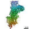
| Title | Structure of Giardia Dicer refined against twinned data | ||||||
 Components Components | GLP_546_48378_50642 | ||||||
 Keywords Keywords | HYDROLASE / Dicer / RNAi / pseudo-merohedral twin | ||||||
| Function / homology |  Function and homology information Function and homology informationHydrolases; Acting on ester bonds; Endoribonucleases producing 5'-phosphomonoesters / ribonuclease III activity / regulatory ncRNA-mediated gene silencing / RNA processing / nucleotide binding / RNA binding / metal ion binding Similarity search - Function | ||||||
| Biological species |  Giardia intestinalis (eukaryote) Giardia intestinalis (eukaryote) | ||||||
| Method |  X-RAY DIFFRACTION / SYNCHROTRON / Resolution: 3 Å X-RAY DIFFRACTION / SYNCHROTRON / Resolution: 3 Å | ||||||
 Authors Authors | Doudna, J.A. / MacRae, I.J. | ||||||
 Citation Citation | Journal: Acta Crystallogr.,Sect.D / Year: 2007 Title: An unusual case of pseudo-merohedral twinning in orthorhombic crystals of Dicer Authors: Macrae, I.J. / Doudna, J.A. | ||||||
| History |
|
- Structure visualization
| Structure viewer | Molecule: MolmilJmol/JSmol |
|---|
- Downloads & links
-Download
| PDBx/mmCIF format | 2qvw.cif.gz | 540.7 KB | Display | PDBx/mmCIF format |
|---|---|---|---|---|
| PDB format | pdb2qvw.ent.gz | 444.9 KB | Display | PDB format |
| PDBx/mmJSON format | 2qvw.json.gz | Tree view | PDBx/mmJSON format | |
| Others |  Other downloads Other downloads |
-Validation report
| Arichive directory | https://data.pdbj.org/pub/pdb/validation_reports/qv/2qvwftp://data.pdbj.org/pub/pdb/validation_reports/qv/2qvw | HTTPS FTP |
|---|
-Related structure data
| Related structure data |  2fflS S: Starting model for refinement |
|---|---|
| Similar structure data |


-Links
 PDBj
PDBj

- Assembly
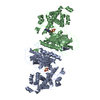
| Deposited unit | 
| ||||||||
|---|---|---|---|---|---|---|---|---|---|
| 1 | 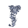
| ||||||||
| 2 | 
| ||||||||
| 3 | 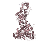
| ||||||||
| 4 | 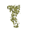
| ||||||||
| Unit cell |
| ||||||||
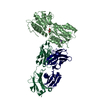
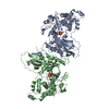
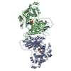
| Details | The biological unit is a monomer. There are four copies in the asymmetric unit. |
-Components
| #1: Protein | Mass: 82690.070 Da / Num. of mol.: 4 Source method: isolated from a genetically manipulated source Source: (gene. exp.) Giardia intestinalis (eukaryote) / Production host:   Spodoptera frugiperda (fall armyworm) Spodoptera frugiperda (fall armyworm)References: UniProt: Q7R2M2, UniProt: A8BQJ3*PLUS, ribonuclease III #2: Chemical | ChemComp-MN /   Mass: 54.938 Da / Num. of mol.: 39 / Source method: obtained synthetically / Formula: Mn Mass: 54.938 Da / Num. of mol.: 39 / Source method: obtained synthetically / Formula: Mn |
|---|
-Experimental details
-Experiment
| Experiment | Method: X-RAY DIFFRACTION / Number of used crystals: 1 |
|---|
- Sample preparation
| Crystal | Density Matthews: 3.17 Å3/Da / Density % sol: 61.17 % |
|---|---|
| Crystal grow | Temperature: 291 K / Method: vapor diffusion, hanging drop / pH: 6.5 Details: 28% PEG 400, 0.1M NaCl, 0.1M MgCl2, 0.1M MES, 5 mM TCEP, pH 6.5, VAPOR DIFFUSION, HANGING DROP, temperature 291.0K |
-Data collection
| Diffraction | Mean temperature: 100 K |
|---|---|
| Diffraction source | Source: SYNCHROTRON / Site: ALS  / Beamline: 8.3.1 / Wavelength: 1 Å / Beamline: 8.3.1 / Wavelength: 1 Å |
| Detector | Type: ADSC QUANTUM 210 / Detector: CCD / Date: May 5, 2005 |
| Radiation | Protocol: SINGLE WAVELENGTH / Monochromatic (M) / Laue (L): M / Scattering type: x-ray |
| Radiation wavelength | Wavelength: 1 Å / Relative weight: 1 |
| Reflection twin | Type: hemihedral / Operator: l,-k,h / Fraction: 0.5 |
| Reflection | Resolution: 3→50 Å / Num. all: 84015 / Num. obs: 82755 / % possible obs: 98.5 % / Observed criterion σ(F): 1 / Observed criterion σ(I): 3 / Redundancy: 5.7 % / Biso Wilson estimate: 68.7 Å2 / Rmerge(I) obs: 0.08 / Rsym value: 0.08 / Net I/σ(I): 20.9 |
| Reflection shell | Resolution: 3→3.11 Å / Redundancy: 5.7 % / Rmerge(I) obs: 0.371 / Mean I/σ(I) obs: 4.21 / Num. unique all: 8238 / Rsym value: 0.371 / % possible all: 99.3 |
- Processing
| Software |
| ||||||||||||||||||||||||||||
|---|---|---|---|---|---|---|---|---|---|---|---|---|---|---|---|---|---|---|---|---|---|---|---|---|---|---|---|---|---|
| Refinement | Starting model: PDB entry 2FFL Resolution: 3→50 Å / σ(F): 1517 Details: The authors state that the structure is the same as the previously deposited Dicer structure (PDB entry 2FFL) except the new model has been refined against higher resolution, twinned data.
| ||||||||||||||||||||||||||||
| Solvent computation | Bsol: 21.105 Å2 | ||||||||||||||||||||||||||||
| Displacement parameters | Biso mean: 39.525 Å2
| ||||||||||||||||||||||||||||
| Refinement step | Cycle: LAST / Resolution: 3→50 Å
| ||||||||||||||||||||||||||||
| Refine LS restraints |
| ||||||||||||||||||||||||||||
| Xplor file |
|
