 Movie
Movie Controller


+ Open data

- Basic information
| Entry | Database: EMDB / ID: EMD-30335 | |||||||||||||||||||||
|---|---|---|---|---|---|---|---|---|---|---|---|---|---|---|---|---|---|---|---|---|---|---|
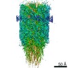
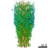
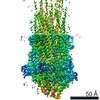
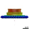
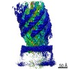
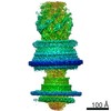
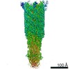
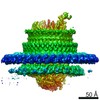
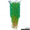
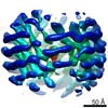
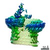
| Title | Cryo-EM structure of the flagellar LP ring from Salmonella | |||||||||||||||||||||
 Map data Map data | Flagellar LP ring from Salmonella | |||||||||||||||||||||
 Sample Sample |
| |||||||||||||||||||||
 Keywords Keywords | Flagella / LP ring / FlgH / FlgI / MOTOR PROTEIN | |||||||||||||||||||||
| Function / homology |  Function and homology information Function and homology informationbacterial-type flagellum basal body, distal rod, L ring / bacterial-type flagellum basal body, distal rod, P ring / cytoskeletal motor activity / bacterial-type flagellum-dependent cell motility / cell outer membrane / outer membrane-bounded periplasmic space / structural molecule activity Similarity search - Function | |||||||||||||||||||||
| Biological species |  Salmonella typhimurium (strain LT2 / SGSC1412 / ATCC 700720) (bacteria) Salmonella typhimurium (strain LT2 / SGSC1412 / ATCC 700720) (bacteria) | |||||||||||||||||||||
| Method | single particle reconstruction / cryo EM / Resolution: 2.8 Å | |||||||||||||||||||||
 Authors Authors | Tan JX / Chang SH | |||||||||||||||||||||
| Funding support |  China, 6 items China, 6 items
| |||||||||||||||||||||
 Citation Citation | Journal: Cell / Year: 2021 Title: Structural basis of assembly and torque transmission of the bacterial flagellar motor. Authors: Jiaxing Tan / Xing Zhang / Xiaofei Wang / Caihuang Xu / Shenghai Chang / Hangjun Wu / Ting Wang / Huihui Liang / Haichun Gao / Yan Zhou / Yongqun Zhu / Abstract: The bacterial flagellar motor is a supramolecular protein machine that drives rotation of the flagellum for motility, which is essential for bacterial survival in different environments and a key ...The bacterial flagellar motor is a supramolecular protein machine that drives rotation of the flagellum for motility, which is essential for bacterial survival in different environments and a key determinant of pathogenicity. The detailed structure of the flagellar motor remains unknown. Here we present an atomic-resolution cryoelectron microscopy (cryo-EM) structure of the bacterial flagellar motor complexed with the hook, consisting of 175 subunits with a molecular mass of approximately 6.3 MDa. The structure reveals that 10 peptides protruding from the MS ring with the FlgB and FliE subunits mediate torque transmission from the MS ring to the rod and overcome the symmetry mismatch between the rotational and helical structures in the motor. The LP ring contacts the distal rod and applies electrostatic forces to support its rotation and torque transmission to the hook. This work provides detailed molecular insights into the structure, assembly, and torque transmission mechanisms of the flagellar motor. | |||||||||||||||||||||
| History |
|
- Structure visualization
| Movie |
Movie viewer |
|---|---|
| Structure viewer | EM map: SurfViewMolmilJmol/JSmol |
| Supplemental images |

- Downloads & links
-EMDB archive
| Map data | emd_30335.map.gz | 56.6 MB | EMDB map data format | |
|---|---|---|---|---|
| Header (meta data) | emd-30335-v30.xmlemd-30335.xml | 13.8 KB 13.8 KB | Display Display | EMDB header |
| Images | 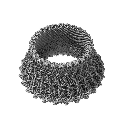 emd_30335.png emd_30335.png | 57.1 KB | ||
| Filedesc metadata | emd-30335.cif.gz | 5.6 KB | ||
| Archive directory |  http://ftp.pdbj.org/pub/emdb/structures/EMD-30335ftp://ftp.pdbj.org/pub/emdb/structures/EMD-30335 http://ftp.pdbj.org/pub/emdb/structures/EMD-30335ftp://ftp.pdbj.org/pub/emdb/structures/EMD-30335 | HTTPS FTP |
-Related structure data
| Related structure data |  7cblMC  7cbmC  7cg0C 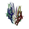 7cg4C  7cg7C 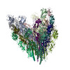 7cgbC 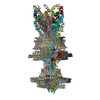 7cgoC 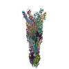 7e80C  7e81C  7e82C M: atomic model generated by this map C: citing same article ( |
|---|---|
| Similar structure data |


-Links
| EMDB pages | EMDB (EBI/PDBe) / EMDataResource |
|---|---|
| Related items in Molecule of the Month |


-Map
| File | Download / File: emd_30335.map.gz / Format: CCP4 / Size: 512 MB / Type: IMAGE STORED AS FLOATING POINT NUMBER (4 BYTES) | ||||||||||||||||||||||||||||||||||||||||||||||||||||||||||||||||||||
|---|---|---|---|---|---|---|---|---|---|---|---|---|---|---|---|---|---|---|---|---|---|---|---|---|---|---|---|---|---|---|---|---|---|---|---|---|---|---|---|---|---|---|---|---|---|---|---|---|---|---|---|---|---|---|---|---|---|---|---|---|---|---|---|---|---|---|---|---|---|
| Annotation | Flagellar LP ring from Salmonella | ||||||||||||||||||||||||||||||||||||||||||||||||||||||||||||||||||||
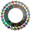
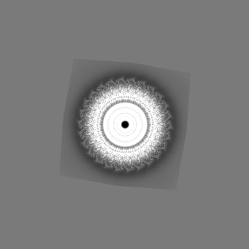
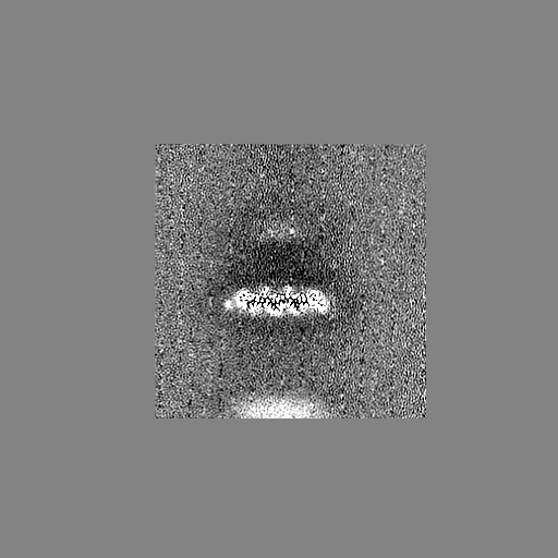
| Projections & slices | Image control
Images are generated by Spider. | ||||||||||||||||||||||||||||||||||||||||||||||||||||||||||||||||||||
| Voxel size | X=Y=Z: 1.307 Å | ||||||||||||||||||||||||||||||||||||||||||||||||||||||||||||||||||||
| Density |
| ||||||||||||||||||||||||||||||||||||||||||||||||||||||||||||||||||||
| Symmetry | Space group: 1 | ||||||||||||||||||||||||||||||||||||||||||||||||||||||||||||||||||||
| Details | EMDB XML:
CCP4 map header:
| ||||||||||||||||||||||||||||||||||||||||||||||||||||||||||||||||||||
 Z (Sec.)
 Y (Row.)
 X (Col.)


-Supplemental data
- Sample components
-Entire : Flagellar hook-basal body
| Entire | Name: Flagellar hook-basal body |
|---|---|
| Components |
|
-Supramolecule #1: Flagellar hook-basal body
| Supramolecule | Name: Flagellar hook-basal body / type: complex / ID: 1 / Parent: 0 / Macromolecule list: #1-#2 |
|---|---|
| Source (natural) | Organism: Salmonella typhimurium (strain LT2 / SGSC1412 / ATCC 700720) (bacteria) |
-Macromolecule #1: Flagellar L-ring protein
| Macromolecule | Name: Flagellar L-ring protein / type: protein_or_peptide / ID: 1 / Number of copies: 26 / Enantiomer: LEVO |
|---|---|
| Source (natural) | Organism: Salmonella typhimurium (strain LT2 / SGSC1412 / ATCC 700720) (bacteria) |
| Molecular weight | Theoretical: 24.726666 KDa |
| Sequence | String: MQKYALHAYP VMALMVATLT GCAWIPAKPL VQGATTAQPI PGPVPVANGS IFQSAQPINY GYQPLFEDRR PRNIGDTLTI VLQENVSAS KSSSANASRD GKTSFGFDTV PRYLQGLFGN SRADMEASGG NSFNGKGGAN ASNTFSGTLT VTVDQVLANG N LHVVGEKQ ...String: MQKYALHAYP VMALMVATLT GCAWIPAKPL VQGATTAQPI PGPVPVANGS IFQSAQPINY GYQPLFEDRR PRNIGDTLTI VLQENVSAS KSSSANASRD GKTSFGFDTV PRYLQGLFGN SRADMEASGG NSFNGKGGAN ASNTFSGTLT VTVDQVLANG N LHVVGEKQ IAINQGTEFI RFSGVVNPRT ISGSNSVPST QVADARIEYV GNGYINEAQN MGWLQRFFLN LSPM UniProtKB: Flagellar L-ring protein |
-Macromolecule #2: Flagellar P-ring protein
| Macromolecule | Name: Flagellar P-ring protein / type: protein_or_peptide / ID: 2 / Number of copies: 26 / Enantiomer: LEVO |
|---|---|
| Source (natural) | Organism: Salmonella typhimurium (strain LT2 / SGSC1412 / ATCC 700720) (bacteria) |
| Molecular weight | Theoretical: 38.194176 KDa |
| Sequence | String: MFKALAGIVL ALVATLAHAE RIRDLTSVQG VRENSLIGYG LVVGLDGTGD QTTQTPFTTQ TLNNMLSQLG ITVPTGTNMQ LKNVAAVMV TASYPPFARQ GQTIDVVVSS MGNAKSLRGG TLLMTPLKGV DSQVYALAQG NILVGGAGAS AGGSSVQVNQ L NGGRITNG ...String: MFKALAGIVL ALVATLAHAE RIRDLTSVQG VRENSLIGYG LVVGLDGTGD QTTQTPFTTQ TLNNMLSQLG ITVPTGTNMQ LKNVAAVMV TASYPPFARQ GQTIDVVVSS MGNAKSLRGG TLLMTPLKGV DSQVYALAQG NILVGGAGAS AGGSSVQVNQ L NGGRITNG AIIERELPTQ FGAGNTINLQ LNDEDFTMAQ QITDAINRAR GYGSATALDA RTVQVRVPSG NSSQVRFLAD IQ NMEVNVT PQDAKVVINS RTGSVVMNRE VTLDSCAVAQ GNLSVTVNRQ LNVNQPNTPF GGGQTVVTPQ TQIDLRQSGG SLQ SVRSSA NLNSVVRALN ALGATPMDLM SILQSMQSAG CLRAKLEII UniProtKB: Flagellar P-ring protein |
-Macromolecule #3: OCTANOIC ACID (CAPRYLIC ACID)
| Macromolecule | Name: OCTANOIC ACID (CAPRYLIC ACID) / type: ligand / ID: 3 / Number of copies: 26 / Formula: OCA |
|---|---|
| Molecular weight | Theoretical: 144.211 Da |
| Chemical component information | 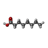 ChemComp-OCA: |
-Experimental details
-Structure determination
| Method | cryo EM |
|---|---|
 Processing Processing | single particle reconstruction |
| Aggregation state | particle |
-Sample preparation
| Buffer | pH: 8 Component:
| ||||||||||||
|---|---|---|---|---|---|---|---|---|---|---|---|---|---|
| Grid | Model: Quantifoil R1.2/1.3 / Material: COPPER / Mesh: 300 / Support film - Material: GRAPHENE OXIDE / Support film - topology: CONTINUOUS / Pretreatment - Type: GLOW DISCHARGE / Pretreatment - Time: 120 sec. | ||||||||||||
| Vitrification | Cryogen name: ETHANE / Chamber humidity: 100 % / Chamber temperature: 295 K / Instrument: FEI VITROBOT MARK IV / Details: blot for 6 seconds before plunging. |
- Electron microscopy
| Microscope | FEI TITAN KRIOS |
|---|---|
| Image recording | Film or detector model: GATAN K2 SUMMIT (4k x 4k) / Detector mode: SUPER-RESOLUTION / Average electron dose: 47.0 e/Å2 |
| Electron beam | Acceleration voltage: 300 kV / Electron source:  FIELD EMISSION GUN FIELD EMISSION GUN |
| Electron optics | Illumination mode: OTHER / Imaging mode: BRIGHT FIELD |
| Experimental equipment |  Model: Titan Krios / Image courtesy: FEI Company |
-Image processing
| Startup model | Type of model: OTHER |
|---|---|
| Final reconstruction | Applied symmetry - Point group: C26 (26 fold cyclic) / Resolution.type: BY AUTHOR / Resolution: 2.8 Å / Resolution method: FSC 0.143 CUT-OFF / Number images used: 80548 |
| Initial angle assignment | Type: MAXIMUM LIKELIHOOD |
| Final angle assignment | Type: MAXIMUM LIKELIHOOD |
-Atomic model buiding 1
| Refinement | Space: REAL / Protocol: AB INITIO MODEL |
|---|---|
| Output model | PDB-7cbl: |
