 Movie
Movie Controller

[English] 日本語
 Yorodumi
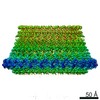
- PDB-6e8g: CryoEM reconstruction of IST1-CHMP1B copolymer filament bound to ... -
+ Open data

- Basic information
| Entry | Database: PDB / ID: 6e8g | |||||||||||||||||||||||||||
|---|---|---|---|---|---|---|---|---|---|---|---|---|---|---|---|---|---|---|---|---|---|---|---|---|---|---|---|---|
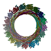
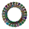
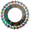
| Title | CryoEM reconstruction of IST1-CHMP1B copolymer filament bound to ssDNA at 2.9 Angstrom resolution | |||||||||||||||||||||||||||
 Components Components |
| |||||||||||||||||||||||||||
 Keywords Keywords | DNA BINDING PROTEIN / PROTEIN FIBRIL / ESCRT-III / CHMP1B / IST1 / ssDNA | |||||||||||||||||||||||||||
| Function / homology |  Function and homology information Function and homology informationMIT domain binding / multivesicular body-lysosome fusion / amphisome membrane / vesicle fusion with vacuole / ESCRT III complex disassembly / late endosome to lysosome transport / ESCRT III complex / kinetochore microtubule / endosome transport via multivesicular body sorting pathway / cytoskeleton-dependent cytokinesis ...MIT domain binding / multivesicular body-lysosome fusion / amphisome membrane / vesicle fusion with vacuole / ESCRT III complex disassembly / late endosome to lysosome transport / ESCRT III complex / kinetochore microtubule / endosome transport via multivesicular body sorting pathway / cytoskeleton-dependent cytokinesis / collateral sprouting / nuclear membrane reassembly / membrane coat / multivesicular body sorting pathway / positive regulation of collateral sprouting / midbody abscission / Sealing of the nuclear envelope (NE) by ESCRT-III / regulation of centrosome duplication / membrane fission / multivesicular body assembly / late endosome to vacuole transport / multivesicular body membrane / ubiquitin-dependent protein catabolic process via the multivesicular body sorting pathway / plasma membrane repair / Flemming body / regulation of mitotic spindle assembly / mitotic metaphase chromosome alignment / nucleus organization / viral budding via host ESCRT complex / endoplasmic reticulum-Golgi intermediate compartment / positive regulation of proteolysis / autophagosome membrane / autophagosome maturation / nuclear pore / multivesicular body / viral budding from plasma membrane / establishment of protein localization / kinetochore / autophagy / azurophil granule lumen / intracellular protein localization / nuclear envelope / protein transport / midbody / endosome membrane / cadherin binding / protein domain specific binding / cell division / lysosomal membrane / Neutrophil degranulation / centrosome / chromatin / protein-containing complex binding / extracellular exosome / extracellular region / nucleoplasm / identical protein binding / plasma membrane / cytosol Similarity search - Function | |||||||||||||||||||||||||||
| Biological species |  Homo sapiens (human) Homo sapiens (human) | |||||||||||||||||||||||||||
| Method | ELECTRON MICROSCOPY / helical reconstruction / cryo EM / Resolution: 2.9 Å | |||||||||||||||||||||||||||
 Authors Authors | Talledge, N. / Frost, A. / McCullough, J. | |||||||||||||||||||||||||||
| Funding support |  United States, 8items United States, 8items
| |||||||||||||||||||||||||||
 Citation Citation | Journal: Biorxiv / Year: 2018 Title: The ESCRT-III proteins IST1 and CHMP1B assemble around nucleic acids Authors: Talledge, N. / McCullough, J. / Wenzel, D.M. / Nguyen, H.C. / Lalonde, M. / Bajorek, M. / Skalicky, J.J. / Frost, A. / Sundquist, W.I. | |||||||||||||||||||||||||||
| History |
|
- Structure visualization
| Movie |
Movie viewer |
|---|---|
| Structure viewer | Molecule: MolmilJmol/JSmol |
- Downloads & links
-Download
| PDBx/mmCIF format | 6e8g.cif.gz | 2.1 MB | Display | PDBx/mmCIF format |
|---|---|---|---|---|
| PDB format | pdb6e8g.ent.gz | Display | PDB format | |
| PDBx/mmJSON format | 6e8g.json.gz | Tree view | PDBx/mmJSON format | |
| Others |  Other downloads Other downloads |
-Validation report
| Arichive directory | https://data.pdbj.org/pub/pdb/validation_reports/e8/6e8gftp://data.pdbj.org/pub/pdb/validation_reports/e8/6e8g | HTTPS FTP |
|---|
-Related structure data
| Related structure data | 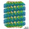 9005MC M: map data used to model this data C: citing same article ( |
|---|---|
| Similar structure data |


-Links
 PDBj
PDBj- Assembly
| Deposited unit | 
|
|---|---|
| 1 |
|
| Symmetry | Helical symmetry: (Circular symmetry: 1 / Dyad axis: no / N subunits divisor: 1 / Num. of operations: 36 / Rise per n subunits: 3.17 Å / Rotation per n subunits: 21.16 °) |
-Components
| #1: Protein | Mass: 40024.711 Da / Num. of mol.: 36 Source method: isolated from a genetically manipulated source Source: (gene. exp.) Homo sapiens (human) / Gene: IST1, KIAA0174 / Plasmid: pET16b / Production host:  #2: Protein | Mass: 22140.354 Da / Num. of mol.: 36 / Mutation: K37E Source method: isolated from a genetically manipulated source Source: (gene. exp.) Homo sapiens (human) / Gene: CHMP1B, C18orf2 / Plasmid: pGEX / Production host: |
|---|
-Experimental details
-Experiment
| Experiment | Method: ELECTRON MICROSCOPY |
|---|---|
| EM experiment | Aggregation state: FILAMENT / 3D reconstruction method: helical reconstruction |
- Sample preparation
| Component | Name: Helical assembly of IST1 and CHMP1B protein bound to ssDNA Type: COMPLEX / Entity ID: all / Source: RECOMBINANT | |||||||||||||||
|---|---|---|---|---|---|---|---|---|---|---|---|---|---|---|---|---|
| Molecular weight | Units: MEGADALTONS / Experimental value: NO | |||||||||||||||
| Source (natural) | Organism: Homo sapiens (human) / Cellular location: cytoplasm | |||||||||||||||
| Source (recombinant) | Organism: | |||||||||||||||
| Buffer solution | pH: 8 | |||||||||||||||
| Buffer component |
| |||||||||||||||
| Specimen | Embedding applied: NO / Shadowing applied: NO / Staining applied: NO / Vitrification applied: YES | |||||||||||||||
| Specimen support | Grid material: COPPER / Grid mesh size: 200 divisions/in. / Grid type: Quantifoil R1.2/1.3 | |||||||||||||||
| Vitrification | Instrument: FEI VITROBOT MARK III / Cryogen name: ETHANE / Humidity: 100 % / Chamber temperature: 292.15 K / Details: 0 mm offset with 10 sec wait time and 2-4 sec blot |
- Electron microscopy imaging
| Experimental equipment |  Model: Titan Krios / Image courtesy: FEI Company |
|---|---|
| Microscopy | Model: FEI TITAN KRIOS |
| Electron gun | Electron source:  FIELD EMISSION GUN / Accelerating voltage: 300 kV / Illumination mode: FLOOD BEAM FIELD EMISSION GUN / Accelerating voltage: 300 kV / Illumination mode: FLOOD BEAM |
| Electron lens | Mode: BRIGHT FIELD / Nominal magnification: 29000 X / Cs: 2.6 mm / C2 aperture diameter: 70 µm / Alignment procedure: COMA FREE |
| Specimen holder | Cryogen: NITROGEN / Specimen holder model: FEI TITAN KRIOS AUTOGRID HOLDER |
| Image recording | Average exposure time: 6.2 sec. / Electron dose: 62 e/Å2 / Detector mode: SUPER-RESOLUTION / Film or detector model: GATAN K2 SUMMIT (4k x 4k) / Num. of grids imaged: 1 / Num. of real images: 2980 |
| Image scans | Width: 3838 / Height: 3710 / Movie frames/image: 32 / Used frames/image: 3-32 |
- Processing
| EM software |
| |||||||||||||||||||||||||||||||||||||||||||||||||||||||
|---|---|---|---|---|---|---|---|---|---|---|---|---|---|---|---|---|---|---|---|---|---|---|---|---|---|---|---|---|---|---|---|---|---|---|---|---|---|---|---|---|---|---|---|---|---|---|---|---|---|---|---|---|---|---|---|---|
| CTF correction | Type: PHASE FLIPPING AND AMPLITUDE CORRECTION | |||||||||||||||||||||||||||||||||||||||||||||||||||||||
| Helical symmerty | Angular rotation/subunit: 21.16 ° / Axial rise/subunit: 3.17 Å / Axial symmetry: C1 | |||||||||||||||||||||||||||||||||||||||||||||||||||||||
| Particle selection | Num. of particles selected: 224252 Details: Helical particle segments were selected using RELION manualpick and extracted using overlapping segments with an asymmetric unit of 17 subunits with a 3.0 Angstrom rise (51 Angstrom total). | |||||||||||||||||||||||||||||||||||||||||||||||||||||||
| 3D reconstruction | Resolution: 2.9 Å / Resolution method: FSC 0.143 CUT-OFF / Num. of particles: 101990 / Num. of class averages: 1 / Symmetry type: HELICAL | |||||||||||||||||||||||||||||||||||||||||||||||||||||||
| Atomic model building | B value: 52.5 / Protocol: AB INITIO MODEL / Space: REAL |
