Movie
Movie Controller

[English] 日本語
 Yorodumi
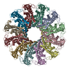
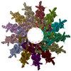
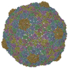
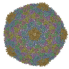
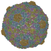
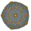
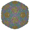
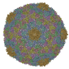
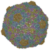
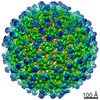
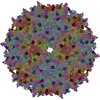
- PDB-2xd8: Capsid structure of the infectious Prochlorococcus Cyanophage P-SSP7 -
+ Open data

- Basic information
| Entry | Database: PDB / ID: 2xd8 | ||||||
|---|---|---|---|---|---|---|---|
| Title | Capsid structure of the infectious Prochlorococcus Cyanophage P-SSP7 | ||||||
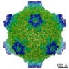
 Components Components | T7-LIKE CAPSID PROTEIN | ||||||
 Keywords Keywords | VIRUS / MARINE PODOVIRUS / T7-LIKE VIRUS | ||||||
| Function / homology | : / Major capsid protein / T7-like capsid protein Function and homology information Function and homology information | ||||||
| Biological species |  PROCHLOROCOCCUS PHAGE P-SSP7 (virus) PROCHLOROCOCCUS PHAGE P-SSP7 (virus) | ||||||
| Method | ELECTRON MICROSCOPY / single particle reconstruction / cryo EM / Resolution: 4.6 Å | ||||||
| Model type details | CA ATOMS ONLY, CHAIN A, B, C, D, E, F, G | ||||||
 Authors Authors | Liu, X. / Zhang, Q. / Murata, K. / Baker, M.L. / Sullivan, M.B. / Fu, C. / Dougherty, M. / Schmid, M.F. / Osburne, M.S. / Chisholm, S.W. / Chiu, W. | ||||||
 Citation Citation | Journal: Nat Struct Mol Biol / Year: 2010 Title: Structural changes in a marine podovirus associated with release of its genome into Prochlorococcus. Authors: Xiangan Liu / Qinfen Zhang / Kazuyoshi Murata / Matthew L Baker / Matthew B Sullivan / Caroline Fu / Matthew T Dougherty / Michael F Schmid / Marcia S Osburne / Sallie W Chisholm / Wah Chiu /  Abstract: Podovirus P-SSP7 infects Prochlorococcus marinus, the most abundant oceanic photosynthetic microorganism. Single-particle cryo-electron microscopy yields icosahedral and asymmetrical structures of ...Podovirus P-SSP7 infects Prochlorococcus marinus, the most abundant oceanic photosynthetic microorganism. Single-particle cryo-electron microscopy yields icosahedral and asymmetrical structures of infectious P-SSP7 with 4.6-A and 9-A resolution, respectively. The asymmetric reconstruction reveals how symmetry mismatches are accommodated among five of the gene products at the portal vertex. Reconstructions of infectious and empty particles show a conformational change of the 'valve' density in the nozzle, an orientation difference in the tail fibers, a disordering of the C terminus of the portal protein and the disappearance of the core proteins. In addition, cryo-electron tomography of P-SSP7 infecting Prochlorococcus showed the same tail-fiber conformation as that in empty particles. Our observations suggest a mechanism whereby, upon binding to the host cell, the tail fibers induce a cascade of structural alterations of the portal vertex complex that triggers DNA release. | ||||||
| History |
|
- Structure visualization
| Movie |
Movie viewer |
|---|---|
| Structure viewer | Molecule: MolmilJmol/JSmol |
- Downloads & links
-Download
| PDBx/mmCIF format | 2xd8.cif.gz | 79 KB | Display | PDBx/mmCIF format |
|---|---|---|---|---|
| PDB format | pdb2xd8.ent.gz | 50.6 KB | Display | PDB format |
| PDBx/mmJSON format | 2xd8.json.gz | Tree view | PDBx/mmJSON format | |
| Others |  Other downloads Other downloads |
-Validation report
| Arichive directory | https://data.pdbj.org/pub/pdb/validation_reports/xd/2xd8ftp://data.pdbj.org/pub/pdb/validation_reports/xd/2xd8 | HTTPS FTP |
|---|
-Related structure data
| Related structure data |  1713MC 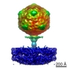 1707C 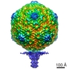 1714C 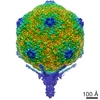 1715C M: map data used to model this data C: citing same article ( |
|---|---|
| Similar structure data |
-Links
 PDBj
PDBj- Assembly
| Deposited unit | 
|
|---|---|
| 1 | x 60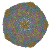
|
| 2 |
|
| 3 | x 5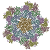
|
| 4 | x 6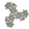
|
| 5 | 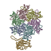
|
| Symmetry | Point symmetry: (Schoenflies symbol: I (icosahedral)) |
-Components
| #1: Protein | Mass: 39494.211 Da / Num. of mol.: 7 Source method: isolated from a genetically manipulated source Source: (gene. exp.) PROCHLOROCOCCUS PHAGE P-SSP7 (virus)Description: P-SSP7 WERE PROPAGATED ON PROCHLOROCOCCUS MED4. Production host:  PROCHLOROCOCCUS MED4 (bacteria) / References: UniProt: Q58N30 PROCHLOROCOCCUS MED4 (bacteria) / References: UniProt: Q58N30 |
|---|
-Experimental details
-Experiment
| Experiment | Method: ELECTRON MICROSCOPY |
|---|---|
| EM experiment | Aggregation state: PARTICLE / 3D reconstruction method: single particle reconstruction |
- Sample preparation
| Component | Name: CYANOPHAGE P-SSP7 / Type: VIRUS |
|---|---|
| Buffer solution | Name: 100 MM TRIS-HCL, 100 MM MGSO4, AND 30 MM NACL / pH: 7.5 / Details: 100 MM TRIS-HCL, 100 MM MGSO4, AND 30 MM NACL |
| Specimen | Conc.: 5 mg/ml / Embedding applied: NO / Shadowing applied: NO / Staining applied: NO / Vitrification applied: YES |
| Specimen support | Details: HOLEY CARBON |
| Vitrification | Instrument: FEI VITROBOT MARK I / Cryogen name: ETHANE |
- Electron microscopy imaging
| Microscopy | Model: JEOL 3200FSC / Date: Aug 31, 2007 |
|---|---|
| Electron gun | Electron source:  FIELD EMISSION GUN / Accelerating voltage: 300 kV / Illumination mode: FLOOD BEAM FIELD EMISSION GUN / Accelerating voltage: 300 kV / Illumination mode: FLOOD BEAM |
| Electron lens | Mode: BRIGHT FIELD / Nominal magnification: 60000 X / Nominal defocus max: 3000 nm / Nominal defocus min: 600 nm / Cs: 4.1 mm |
| Specimen holder | Temperature: 101 K |
| Image recording | Electron dose: 20 e/Å2 / Film or detector model: KODAK SO-163 FILM |
| Image scans | Num. digital images: 1059 |
| Radiation wavelength | Relative weight: 1 |
- Processing
| EM software | Name: MPSA / Category: 3D reconstruction | |||||||||||||||||||||
|---|---|---|---|---|---|---|---|---|---|---|---|---|---|---|---|---|---|---|---|---|---|---|
| CTF correction | Details: INDIVIDUAL MICROGRAPH | |||||||||||||||||||||
| Symmetry | Point symmetry: I (icosahedral) | |||||||||||||||||||||
| 3D reconstruction | Method: CROSS-COMMON LINES / Resolution: 4.6 Å / Num. of particles: 36000 / Nominal pixel size: 1.27 Å / Actual pixel size: 1.17 Å / Magnification calibration: 2D CRYSTAL Details: SUBMISSION BASED ON EXPERIMENTAL DATA FROM EMDB EMD-1713. Symmetry type: POINT | |||||||||||||||||||||
| Atomic model building |
| |||||||||||||||||||||
| Refinement | Highest resolution: 4.6 Å | |||||||||||||||||||||
| Refinement step | Cycle: LAST / Highest resolution: 4.6 Å
|