Movie
Movie Controller

[English] 日本語
 Yorodumi
- EMDB-1713: Capsid structure of the infectious Prochlorococcus Cyanophage P-SSP7 -
+ Open data

- Basic information
| Entry | Database: EMDB / ID: EMD-1713 | |||||||||
|---|---|---|---|---|---|---|---|---|---|---|
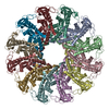
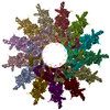
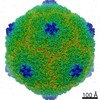
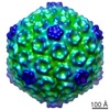
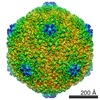
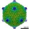
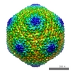
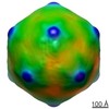
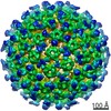
| Title | Capsid structure of the infectious Prochlorococcus Cyanophage P-SSP7 | |||||||||
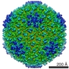
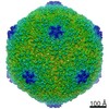
 Map data Map data | Icosahedral capsid structure of cyanophage P-SSP7 | |||||||||
 Sample Sample |
| |||||||||
 Keywords Keywords | Marine Podovirus / T7-like virus / infects Prochlorococcus MED4 | |||||||||
| Function / homology | : / Major capsid protein / T7-like capsid protein Function and homology information Function and homology information | |||||||||
| Biological species |  Prochlorococcus phage P-SSP7 (virus) Prochlorococcus phage P-SSP7 (virus) | |||||||||
| Method | single particle reconstruction / cryo EM / negative staining / Resolution: 4.6 Å | |||||||||
 Authors Authors | Liu X / Zhang Q / Murata K / Baker ML / Sullivan MB / Fu C / Dougherty M / Schmid MF / Osburne MS / Chisholm SW / Chiu W | |||||||||
 Citation Citation | Journal: Nat Struct Mol Biol / Year: 2010 Title: Structural changes in a marine podovirus associated with release of its genome into Prochlorococcus. Authors: Xiangan Liu / Qinfen Zhang / Kazuyoshi Murata / Matthew L Baker / Matthew B Sullivan / Caroline Fu / Matthew T Dougherty / Michael F Schmid / Marcia S Osburne / Sallie W Chisholm / Wah Chiu /  Abstract: Podovirus P-SSP7 infects Prochlorococcus marinus, the most abundant oceanic photosynthetic microorganism. Single-particle cryo-electron microscopy yields icosahedral and asymmetrical structures of ...Podovirus P-SSP7 infects Prochlorococcus marinus, the most abundant oceanic photosynthetic microorganism. Single-particle cryo-electron microscopy yields icosahedral and asymmetrical structures of infectious P-SSP7 with 4.6-A and 9-A resolution, respectively. The asymmetric reconstruction reveals how symmetry mismatches are accommodated among five of the gene products at the portal vertex. Reconstructions of infectious and empty particles show a conformational change of the 'valve' density in the nozzle, an orientation difference in the tail fibers, a disordering of the C terminus of the portal protein and the disappearance of the core proteins. In addition, cryo-electron tomography of P-SSP7 infecting Prochlorococcus showed the same tail-fiber conformation as that in empty particles. Our observations suggest a mechanism whereby, upon binding to the host cell, the tail fibers induce a cascade of structural alterations of the portal vertex complex that triggers DNA release. | |||||||||
| History |
|
- Structure visualization
| Movie |
Movie viewer |
|---|---|
| Structure viewer | EM map: SurfViewMolmilJmol/JSmol |
| Supplemental images |
- Downloads & links
-EMDB archive
| Map data | emd_1713.map.gz | 97.9 MB | EMDB map data format | |
|---|---|---|---|---|
| Header (meta data) | emd-1713-v30.xmlemd-1713.xml | 12.1 KB 12.1 KB | Display Display | EMDB header |
| Images | 412.9 KB | |||
| Archive directory |  http://ftp.pdbj.org/pub/emdb/structures/EMD-1713ftp://ftp.pdbj.org/pub/emdb/structures/EMD-1713 http://ftp.pdbj.org/pub/emdb/structures/EMD-1713ftp://ftp.pdbj.org/pub/emdb/structures/EMD-1713 | HTTPS FTP |
-Related structure data
| Related structure data | 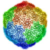 2xd8MC 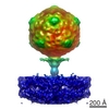 1707C 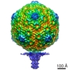 1714C 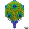 1715C C: citing same article ( M: atomic model generated by this map |
|---|---|
| Similar structure data |

-Links
| EMDB pages | EMDB (EBI/PDBe) / EMDataResource |
|---|
-Map
| File | Download / File: emd_1713.map.gz / Format: CCP4 / Size: 711.9 MB / Type: IMAGE STORED AS FLOATING POINT NUMBER (4 BYTES) | ||||||||||||||||||||||||||||||||||||||||||||||||||||||||||||||||||||
|---|---|---|---|---|---|---|---|---|---|---|---|---|---|---|---|---|---|---|---|---|---|---|---|---|---|---|---|---|---|---|---|---|---|---|---|---|---|---|---|---|---|---|---|---|---|---|---|---|---|---|---|---|---|---|---|---|---|---|---|---|---|---|---|---|---|---|---|---|---|
| Annotation | Icosahedral capsid structure of cyanophage P-SSP7 | ||||||||||||||||||||||||||||||||||||||||||||||||||||||||||||||||||||
| Projections & slices | Image control
Images are generated by Spider. | ||||||||||||||||||||||||||||||||||||||||||||||||||||||||||||||||||||
| Voxel size | X=Y=Z: 1.17 Å | ||||||||||||||||||||||||||||||||||||||||||||||||||||||||||||||||||||
| Density |
| ||||||||||||||||||||||||||||||||||||||||||||||||||||||||||||||||||||
| Symmetry | Space group: 1 | ||||||||||||||||||||||||||||||||||||||||||||||||||||||||||||||||||||
| Details | EMDB XML:
CCP4 map header:
| ||||||||||||||||||||||||||||||||||||||||||||||||||||||||||||||||||||
 Z (Sec.)
 Y (Row.)
 X (Col.)


-Supplemental data
- Sample components
-Entire : Cyanophage P-SSP7
| Entire | Name: Cyanophage P-SSP7 |
|---|---|
| Components |
|
-Supramolecule #1000: Cyanophage P-SSP7
| Supramolecule | Name: Cyanophage P-SSP7 / type: sample / ID: 1000 / Number unique components: 1 |
|---|
-Supramolecule #1: Prochlorococcus phage P-SSP7
| Supramolecule | Name: Prochlorococcus phage P-SSP7 / type: virus / ID: 1 / Name.synonym: T7-LIKE CAPSID Details: The gp10 protein of P-SSP7 contains 375 amino acids. NCBI-ID: 268748 / Sci species name: Prochlorococcus phage P-SSP7 / Virus type: VIRION / Virus isolate: STRAIN / Virus enveloped: No / Virus empty: No / Syn species name: T7-LIKE CAPSID |
|---|---|
| Host (natural) | Organism:  Prochlorococcus (bacteria) / synonym: BACTERIA(EUBACTERIA) Prochlorococcus (bacteria) / synonym: BACTERIA(EUBACTERIA) |
| Virus shell | Shell ID: 1 / Diameter: 655 Å / T number (triangulation number): 7 |
-Experimental details
-Structure determination
| Method | negative staining, cryo EM |
|---|---|
 Processing Processing | single particle reconstruction |
| Aggregation state | particle |
-Sample preparation
| Concentration | 5 mg/mL |
|---|---|
| Buffer | pH: 7.5 / Details: 100 mM Tris-HCl , 100 mM MgSO4, and 30 mM NaCl |
| Staining | Type: NEGATIVE / Details: No stain |
| Grid | Details: 200 mesh copper grid |
| Vitrification | Cryogen name: ETHANE / Chamber humidity: 30 % / Chamber temperature: 101 K / Instrument: OTHER / Details: Vitrification instrument: FEI Vitrobot / Method: Blot for 2 seconds 2 times before plunging |
- Electron microscopy
| Microscope | JEOL 3200FSC |
|---|---|
| Temperature | Min: 4.5 K / Max: 102 K / Average: 101 K |
| Alignment procedure | Legacy - Astigmatism: Objective lens astigmatism was corrected at 400,000 times magnification |
| Specialist optics | Energy filter - Name: JEOL / Energy filter - Lower energy threshold: 15.0 eV / Energy filter - Upper energy threshold: 20.0 eV |
| Details | MDS |
| Date | Aug 31, 2007 |
| Image recording | Category: FILM / Film or detector model: KODAK SO-163 FILM / Digitization - Scanner: NIKON SUPER COOLSCAN 9000 / Digitization - Sampling interval: 6.35 µm / Number real images: 1059 / Average electron dose: 20 e/Å2 Details: The cryoEM images were recorded on Kodak SO163 films. The films were developed in full strength D19 Kodak developer for 12 minutes at 20 degree and fixed for 10 minutes in Kodak fixer. The ...Details: The cryoEM images were recorded on Kodak SO163 films. The films were developed in full strength D19 Kodak developer for 12 minutes at 20 degree and fixed for 10 minutes in Kodak fixer. The films were digitized at 6.35 microns per pixel using a Nikon Super CoolScan 9000 ED scanner (Nikon Corp., Japan). Bits/pixel: 8 |
| Electron beam | Acceleration voltage: 300 kV / Electron source:  FIELD EMISSION GUN FIELD EMISSION GUN |
| Electron optics | Illumination mode: SPOT SCAN / Imaging mode: BRIGHT FIELD / Cs: 4.1 mm / Nominal defocus max: 3.0 µm / Nominal defocus min: 0.6 µm / Nominal magnification: 60000 |
| Sample stage | Specimen holder: Side entry / Specimen holder model: JEOL 3200FSC CRYOHOLDER |
-Image processing
| Details | The particle were selected by the consistency criterion of MPSA |
|---|---|
| CTF correction | Details: Each Micrograph |
| Final reconstruction | Applied symmetry - Point group: I (icosahedral) / Algorithm: OTHER / Resolution.type: BY AUTHOR / Resolution: 4.6 Å / Resolution method: FSC 0.5 CUT-OFF / Software - Name: MPSA Details: The map was directly built from raw particles with icosahedral symmetry enforced. Number images used: 36000 |
| Final angle assignment | Details: EMAN: Z along 5-fold and Y along 2-fold axis. |
-Atomic model buiding 1
| Initial model | PDB ID: |
|---|---|
| Refinement | Space: REAL |
| Output model | PDB-2xd8: |