 Movie
Movie Controller

[English] 日本語
 Yorodumi
- EMDB-1707: Structural Changes in a Marine Podovirus Associated with Viral Ge... -
+ Open data

- Basic information
| Entry | Database: EMDB / ID: EMD-1707 | |||||||||
|---|---|---|---|---|---|---|---|---|---|---|
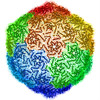
| Title | Structural Changes in a Marine Podovirus Associated with Viral Genome Release into Prochlorococcus | |||||||||
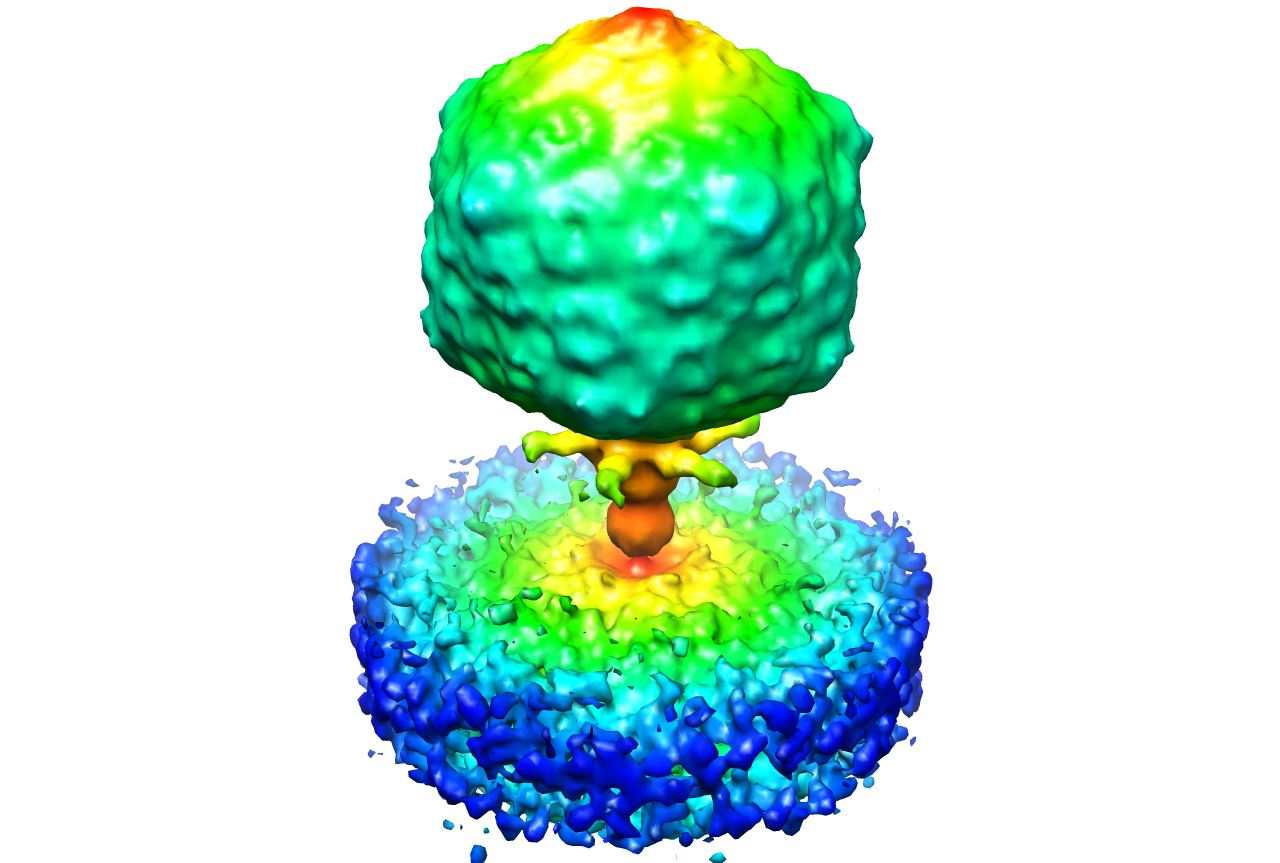
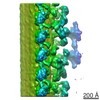
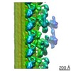
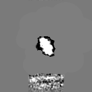
 Map data Map data | This is an image of a surface rendered top-view of P-SSP7 on the host cell surface | |||||||||
 Sample Sample |
| |||||||||
 Keywords Keywords | Viral infection / electron cryo-tomography | |||||||||
| Biological species |  Prochlorococcus phage P-SSP7 (virus) Prochlorococcus phage P-SSP7 (virus) | |||||||||
| Method | subtomogram averaging / cryo EM / negative staining | |||||||||
 Authors Authors | Liu X / Zhang Q / Murata K / Baker ML / Sullivan MB / Fu C / Dougherty M / Schmid MF / Osburne MS / Chisholm SW / Chiu W | |||||||||
 Citation Citation | Journal: Nat Struct Mol Biol / Year: 2010 Title: Structural changes in a marine podovirus associated with release of its genome into Prochlorococcus. Authors: Xiangan Liu / Qinfen Zhang / Kazuyoshi Murata / Matthew L Baker / Matthew B Sullivan / Caroline Fu / Matthew T Dougherty / Michael F Schmid / Marcia S Osburne / Sallie W Chisholm / Wah Chiu /  Abstract: Podovirus P-SSP7 infects Prochlorococcus marinus, the most abundant oceanic photosynthetic microorganism. Single-particle cryo-electron microscopy yields icosahedral and asymmetrical structures of ...Podovirus P-SSP7 infects Prochlorococcus marinus, the most abundant oceanic photosynthetic microorganism. Single-particle cryo-electron microscopy yields icosahedral and asymmetrical structures of infectious P-SSP7 with 4.6-A and 9-A resolution, respectively. The asymmetric reconstruction reveals how symmetry mismatches are accommodated among five of the gene products at the portal vertex. Reconstructions of infectious and empty particles show a conformational change of the 'valve' density in the nozzle, an orientation difference in the tail fibers, a disordering of the C terminus of the portal protein and the disappearance of the core proteins. In addition, cryo-electron tomography of P-SSP7 infecting Prochlorococcus showed the same tail-fiber conformation as that in empty particles. Our observations suggest a mechanism whereby, upon binding to the host cell, the tail fibers induce a cascade of structural alterations of the portal vertex complex that triggers DNA release. | |||||||||
| History |
|
- Structure visualization
| Movie |
 Movie viewer Movie viewer |
|---|---|
| Structure viewer | EM map: SurfViewMolmilJmol/JSmol |
| Supplemental images |
- Downloads & links
-EMDB archive
| Map data | emd_1707.map.gz | 6.5 MB | EMDB map data format | |
|---|---|---|---|---|
| Header (meta data) | emd-1707-v30.xmlemd-1707.xml | 11.3 KB 11.3 KB | Display Display | EMDB header |
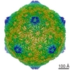
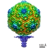
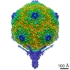
| Images |  1707.png 1707.png | 547.1 KB | ||
| Archive directory |  http://ftp.pdbj.org/pub/emdb/structures/EMD-1707ftp://ftp.pdbj.org/pub/emdb/structures/EMD-1707 http://ftp.pdbj.org/pub/emdb/structures/EMD-1707ftp://ftp.pdbj.org/pub/emdb/structures/EMD-1707 | HTTPS FTP |
-Related structure data
-Links
| EMDB pages | EMDB (EBI/PDBe) / EMDataResource |
|---|
-Map
| File | Download / File: emd_1707.map.gz / Format: CCP4 / Size: 21.7 MB / Type: IMAGE STORED AS FLOATING POINT NUMBER (4 BYTES) | ||||||||||||||||||||||||||||||||||||||||||||||||||||||||||||||||||||
|---|---|---|---|---|---|---|---|---|---|---|---|---|---|---|---|---|---|---|---|---|---|---|---|---|---|---|---|---|---|---|---|---|---|---|---|---|---|---|---|---|---|---|---|---|---|---|---|---|---|---|---|---|---|---|---|---|---|---|---|---|---|---|---|---|---|---|---|---|---|
| Annotation | This is an image of a surface rendered top-view of P-SSP7 on the host cell surface | ||||||||||||||||||||||||||||||||||||||||||||||||||||||||||||||||||||
| Projections & slices | Image control
Images are generated by Spider. | ||||||||||||||||||||||||||||||||||||||||||||||||||||||||||||||||||||
| Voxel size | X=Y=Z: 9.36 Å | ||||||||||||||||||||||||||||||||||||||||||||||||||||||||||||||||||||
| Density |
| ||||||||||||||||||||||||||||||||||||||||||||||||||||||||||||||||||||
| Symmetry | Space group: 1 | ||||||||||||||||||||||||||||||||||||||||||||||||||||||||||||||||||||
| Details | EMDB XML:
CCP4 map header:
| ||||||||||||||||||||||||||||||||||||||||||||||||||||||||||||||||||||
 Z (Sec.)
 Y (Row.)
 X (Col.)


-Supplemental data
- Sample components
-Entire : P-SSP7 of Prochlorococcus
| Entire | Name: P-SSP7 of Prochlorococcus |
|---|---|
| Components |
|
-Supramolecule #1000: P-SSP7 of Prochlorococcus
| Supramolecule | Name: P-SSP7 of Prochlorococcus / type: sample / ID: 1000 / Number unique components: 1 |
|---|
-Supramolecule #1: Prochlorococcus phage P-SSP7
| Supramolecule | Name: Prochlorococcus phage P-SSP7 / type: virus / ID: 1 / Name.synonym: P-SSP7 / NCBI-ID: 268748 / Sci species name: Prochlorococcus phage P-SSP7 / Database: NCBI / Virus type: VIRION / Virus isolate: STRAIN / Virus enveloped: No / Virus empty: No / Syn species name: P-SSP7 |
|---|---|
| Host (natural) | Organism: Podoviridae / synonym: BACTERIA(EUBACTERIA) |
| Virus shell | Shell ID: 1 / Diameter: 655 Å / T number (triangulation number): 7 |
-Experimental details
-Structure determination
| Method | negative staining, cryo EM |
|---|---|
 Processing Processing | subtomogram averaging |
| Aggregation state | particle |
-Sample preparation
| Buffer | pH: 7.5 Details: 100 mM Tris-HCl (pH 7.5), 100 mM MgSO4, and 30 mM NaCl |
|---|---|
| Staining | Type: NEGATIVE Details: Grids with sample solution were quickly frozen in liquid ethane. |
| Grid | Details: R3.5/1 Quantifoil |
| Vitrification | Cryogen name: ETHANE / Chamber humidity: 40 % / Chamber temperature: 90 K / Instrument: LEICA EM CPC / Details: Vitrification instrument: Leica CPC / Method: Blot for 2 seconds before plunging |
- Electron microscopy
| Microscope | JEOL 3200FSC |
|---|---|
| Temperature | Min: 90 K / Max: 95 K / Average: 90 K |
| Alignment procedure | Legacy - Astigmatism: Objective lens astigmatism was corrected at 100,000 times magnification |
| Specialist optics | Energy filter - Name: Omega / Energy filter - Lower energy threshold: 0.0 eV / Energy filter - Upper energy threshold: 30.0 eV |
| Image recording | Category: CCD / Film or detector model: GATAN ULTRASCAN 4000 (4k x 4k) / Digitization - Sampling interval: 15 µm / Average electron dose: 80 e/Å2 / Bits/pixel: 16 |
| Electron beam | Acceleration voltage: 300 kV / Electron source:  FIELD EMISSION GUN FIELD EMISSION GUN |
| Electron optics | Calibrated magnification: 30000 / Illumination mode: FLOOD BEAM / Imaging mode: BRIGHT FIELD / Cs: 4.1 mm / Nominal defocus max: 5.0 µm / Nominal defocus min: 3.0 µm / Nominal magnification: 20000 |
| Sample stage | Specimen holder: Eucentric / Specimen holder model: JEOL 3200FSC CRYOHOLDER / Tilt series - Axis1 - Min angle: -62 ° / Tilt series - Axis1 - Max angle: 62 ° |
-Image processing
| Final reconstruction | Algorithm: OTHER / Software - Name: IMOD |
|---|
