 Movie
Movie Controller

[English] 日本語
 Yorodumi
- PDB-2des: INTERACTIONS BETWEEN MORPHOLINYL ANTHRACYCLINES AND DNA: THE CRYS... -
+ Open data

- Basic information
| Entry | Database: PDB / ID: 2des | ||||||||||||||||||
|---|---|---|---|---|---|---|---|---|---|---|---|---|---|---|---|---|---|---|---|
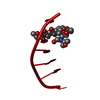
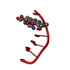
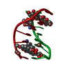
| Title | INTERACTIONS BETWEEN MORPHOLINYL ANTHRACYCLINES AND DNA: THE CRYSTAL STRUCTURE OF A MORPHOLINO DOXORUBICIN BOUND TO D(CGTACG) | ||||||||||||||||||
 Components Components | DNA (5'-D(* Keywords KeywordsDNA / RIGHT HANDED DNA / DOUBLE HELIX / COMPLEXED WITH DRUG | Function / homology | Chem-DMM / DNA |  Function and homology information Function and homology informationMethod |  X-RAY DIFFRACTION / Resolution: 1.5 Å X-RAY DIFFRACTION / Resolution: 1.5 Å  Authors AuthorsCirilli, M. / Bachechi, F. / Ughetto, G. / Colonna, F.P. / Capobianco, M.L. |  CitationJournal: J.Mol.Biol. / Year: 1993 CitationJournal: J.Mol.Biol. / Year: 1993Title: Interactions between morpholinyl anthracyclines and DNA. The crystal structure of a morpholino doxorubicin bound to d(CGTACG). Authors: Cirilli, M. / Bachechi, F. / Ughetto, G. / Colonna, F.P. / Capobianco, M.L. #1: Journal: Biochemistry / Year: 1987Title: Interactions Between an Anthracycline Antibiotic and DNA: Molecular Structure of Daunomycin Complexed to d(CpGpTpApCpG) at 1.2-Angstroms Resolution Authors: Wang, A.-H.J. / Ughetto, G. / Quigley, G.J. / Rich, A. #2: Journal: J.Mol.Biol. / Year: 1990Title: Structure of 11-Deoxydaunomycin Bound to DNA Containing a Phosphorothioate Authors: Williams, L.D. / Egli, M. / Ughetto, G. / Van Der Marel, G.A. / Van Boom, J.H. / Quigley, G.J. / Wang, A.H.-J. / Rich, A. / Frederick, C.A. History |
|
- Structure visualization
| Structure viewer | Molecule: MolmilJmol/JSmol |
|---|
- Downloads & links
-Download
| PDBx/mmCIF format | 2des.cif.gz | 24.8 KB | Display | PDBx/mmCIF format |
|---|---|---|---|---|
| PDB format | pdb2des.ent.gz | 14.7 KB | Display | PDB format |
| PDBx/mmJSON format | 2des.json.gz | Tree view | PDBx/mmJSON format | |
| Others |  Other downloads Other downloads |
-Validation report
| Arichive directory | https://data.pdbj.org/pub/pdb/validation_reports/de/2desftp://data.pdbj.org/pub/pdb/validation_reports/de/2des | HTTPS FTP |
|---|
-Related structure data
| Similar structure data |
|---|


-Links
 PDBj
PDBj

- Assembly
| Deposited unit | 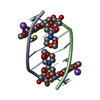
| ||||||||
|---|---|---|---|---|---|---|---|---|---|
| 1 |
| ||||||||
| Unit cell |
|
-Components
| #1: DNA chain | Mass: 1809.217 Da / Num. of mol.: 2 / Source method: obtained synthetically #2: Chemical |   Mass: 643.635 Da / Num. of mol.: 2 / Source method: obtained synthetically / Formula: C32H37NO13 Mass: 643.635 Da / Num. of mol.: 2 / Source method: obtained synthetically / Formula: C32H37NO13#3: Chemical |   Mass: 22.990 Da / Num. of mol.: 2 / Source method: obtained synthetically / Formula: Na Mass: 22.990 Da / Num. of mol.: 2 / Source method: obtained synthetically / Formula: Na#4: Chemical | ChemComp-MG / |   Mass: 24.305 Da / Num. of mol.: 1 / Source method: obtained synthetically / Formula: Mg Mass: 24.305 Da / Num. of mol.: 1 / Source method: obtained synthetically / Formula: Mg#5: Water | ChemComp-HOH / |  Mass: 18.015 Da / Num. of mol.: 79 / Source method: isolated from a natural source / Formula: H2O Mass: 18.015 Da / Num. of mol.: 79 / Source method: isolated from a natural source / Formula: H2O |
|---|
-Experimental details
-Experiment
| Experiment | Method: X-RAY DIFFRACTION |
|---|
- Sample preparation
| Crystal | Density Matthews: 2.45 Å3/Da / Density % sol: 49.82 % | ||||||||||||||||||||||||||||||||
|---|---|---|---|---|---|---|---|---|---|---|---|---|---|---|---|---|---|---|---|---|---|---|---|---|---|---|---|---|---|---|---|---|---|
| Crystal grow | Method: vapor diffusion, sitting drop / pH: 5.5 / Details: pH 5.50, VAPOR DIFFUSION, SITTING DROP / Temp details: ROOM TEMPERATURE | ||||||||||||||||||||||||||||||||
| Components of the solutions |
| ||||||||||||||||||||||||||||||||
| Crystal grow | *PLUS pH: 5.5 / Method: other | ||||||||||||||||||||||||||||||||
| Components of the solutions | *PLUS
|
-Data collection
| Diffraction | Ambient temp details: ROOM TEMPERATURE |
|---|---|
| Detector | Type: SYNTEX P21 / Detector: DIFFRACTOMETER |
| Radiation | Scattering type: x-ray |
| Radiation wavelength | Relative weight: 1 |
| Reflection | Num. obs: 3988 |
| Reflection | *PLUS |
- Processing
| Software | Name: NUCLSQ / Classification: refinement | ||||||||||||
|---|---|---|---|---|---|---|---|---|---|---|---|---|---|
| Refinement | Resolution: 1.5→10 Å / σ(F): 2 /
| ||||||||||||
| Refine Biso |
| ||||||||||||
| Refinement step | Cycle: LAST / Resolution: 1.5→10 Å
| ||||||||||||
| Refinement | *PLUS Highest resolution: 1.5 Å / Lowest resolution: 10 Å / Num. reflection obs: 3588 / Rfactor obs: 0.192 / σ(F): 2 | ||||||||||||
| Solvent computation | *PLUS | ||||||||||||
| Displacement parameters | *PLUS |
