 Movie
Movie Controller

[English] 日本語
 Yorodumi
- PDB-234d: CRYSTAL STRUCTURE OF FOUR MORPHOLINO-DOXORUBICIN ANTICANCER DRUGS... -
+ Open data

- Basic information
| Entry | Database: PDB / ID: 234d | ||||||||||||||||||
|---|---|---|---|---|---|---|---|---|---|---|---|---|---|---|---|---|---|---|---|
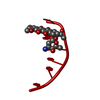
| Title | CRYSTAL STRUCTURE OF FOUR MORPHOLINO-DOXORUBICIN ANTICANCER DRUGS COMPLEXED WITH D(CGTACG) AND D(CGATCG): IMPLICATIONS IN DRUG-DNA CROSSLINK | ||||||||||||||||||
 Components Components | DNA (5'-D(* Keywords KeywordsDNA / RIGHT HANDED DNA / DOUBLE HELIX / COMPLEXED WITH DRUG | Function / homology | Chem-DMM / DNA |  Function and homology information Function and homology informationMethod |  X-RAY DIFFRACTION / Resolution: 1.8 Å X-RAY DIFFRACTION / Resolution: 1.8 Å  Authors AuthorsGao, Y.-G. / Wang, A.H.-J. |  CitationJournal: J.Biomol.Struct.Dyn. / Year: 1995 CitationJournal: J.Biomol.Struct.Dyn. / Year: 1995Title: Crystal structures of four morpholino-doxorubicin anticancer drugs complexed with d(CGTACG) and d(CGATCG): implications in drug-DNA crosslink. Authors: Gao, Y.G. / Wang, A.H. History |
|
- Structure visualization
| Structure viewer | Molecule: MolmilJmol/JSmol |
|---|
- Downloads & links
-Download
| PDBx/mmCIF format | 234d.cif.gz | 15.3 KB | Display | PDBx/mmCIF format |
|---|---|---|---|---|
| PDB format | pdb234d.ent.gz | 8.5 KB | Display | PDB format |
| PDBx/mmJSON format | 234d.json.gz | Tree view | PDBx/mmJSON format | |
| Others |  Other downloads Other downloads |
-Validation report
| Arichive directory | https://data.pdbj.org/pub/pdb/validation_reports/34/234dftp://data.pdbj.org/pub/pdb/validation_reports/34/234d | HTTPS FTP |
|---|
-Related structure data


-Links
 PDBj
PDBj

- Assembly
| Deposited unit | 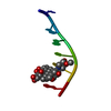
| ||||||||
|---|---|---|---|---|---|---|---|---|---|
| 1 | 
| ||||||||
| Unit cell |
|
-Components
| #1: DNA chain | Mass: 1809.217 Da / Num. of mol.: 1 / Source method: obtained synthetically |
|---|---|
| #2: Chemical | ChemComp-DMM /   Mass: 643.635 Da / Num. of mol.: 1 / Source method: obtained synthetically / Formula: C32H37NO13 Mass: 643.635 Da / Num. of mol.: 1 / Source method: obtained synthetically / Formula: C32H37NO13 |
| #3: Water | ChemComp-HOH /  Mass: 18.015 Da / Num. of mol.: 49 / Source method: isolated from a natural source / Formula: H2O Mass: 18.015 Da / Num. of mol.: 49 / Source method: isolated from a natural source / Formula: H2O |
-Experimental details
-Experiment
| Experiment | Method: X-RAY DIFFRACTION |
|---|
- Sample preparation
| Crystal | Density Matthews: 2.95 Å3/Da / Density % sol: 58.29 % | ||||||||||||||||||||||||||||||||||||||||||||||||||||||||
|---|---|---|---|---|---|---|---|---|---|---|---|---|---|---|---|---|---|---|---|---|---|---|---|---|---|---|---|---|---|---|---|---|---|---|---|---|---|---|---|---|---|---|---|---|---|---|---|---|---|---|---|---|---|---|---|---|---|
| Crystal grow | Method: vapor diffusion / pH: 6 / Details: pH 6.00, VAPOR DIFFUSION / Temp details: ROOM TEMPERATURE | ||||||||||||||||||||||||||||||||||||||||||||||||||||||||
| Components of the solutions |
| ||||||||||||||||||||||||||||||||||||||||||||||||||||||||
| Crystal grow | *PLUS pH: 6 | ||||||||||||||||||||||||||||||||||||||||||||||||||||||||
| Components of the solutions | *PLUS
|
-Data collection
| Diffraction | Mean temperature: 298 K |
|---|---|
| Diffraction source | Source: ROTATING ANODE / Type: RIGAKU / Wavelength: 1.5418 |
| Detector | Type: RIGAKU AFC-5 / Detector: DIFFRACTOMETER |
| Radiation | Scattering type: x-ray |
| Radiation wavelength | Wavelength: 1.5418 Å / Relative weight: 1 |
| Reflection | Highest resolution: 1.7 Å |
| Reflection | *PLUS Highest resolution: 1.7 Å / Num. obs: 1327 |
- Processing
| Software | Name: NUCLSQ / Classification: refinement | |||||||||||||||||||||||||||||||||||||||||||||||||||||||||||||||
|---|---|---|---|---|---|---|---|---|---|---|---|---|---|---|---|---|---|---|---|---|---|---|---|---|---|---|---|---|---|---|---|---|---|---|---|---|---|---|---|---|---|---|---|---|---|---|---|---|---|---|---|---|---|---|---|---|---|---|---|---|---|---|---|---|
| Refinement | Highest resolution: 1.8 Å / σ(F): 2 /
| |||||||||||||||||||||||||||||||||||||||||||||||||||||||||||||||
| Refinement step | Cycle: LAST / Highest resolution: 1.8 Å
| |||||||||||||||||||||||||||||||||||||||||||||||||||||||||||||||
| Refine LS restraints |
| |||||||||||||||||||||||||||||||||||||||||||||||||||||||||||||||
| Refinement | *PLUS Highest resolution: 1.8 Å / σ(F): 2 / Rfactor obs: 0.19 | |||||||||||||||||||||||||||||||||||||||||||||||||||||||||||||||
| Solvent computation | *PLUS | |||||||||||||||||||||||||||||||||||||||||||||||||||||||||||||||
| Displacement parameters | *PLUS | |||||||||||||||||||||||||||||||||||||||||||||||||||||||||||||||
| Refine LS restraints | *PLUS Type: n_bond_d / Dev ideal: 0.018 |
