 Movie
Movie Controller

[English] 日本語
 Yorodumi
- PDB-1p20: Surprising Roles of Electrostatic Interactions in DNA-Ligand Complexes -
+ Open data

- Basic information
| Entry | Database: PDB / ID: 1p20 | ||||||||||||||||||
|---|---|---|---|---|---|---|---|---|---|---|---|---|---|---|---|---|---|---|---|
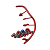
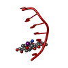
| Title | Surprising Roles of Electrostatic Interactions in DNA-Ligand Complexes | ||||||||||||||||||
 Components Components | 5'-D(* Keywords KeywordsDNA / adriamycin / intercalation / electrostatics / thallium | Function / homology | DOXORUBICIN / THALLIUM (I) ION / DNA |  Function and homology information Function and homology informationMethod |  X-RAY DIFFRACTION / MOLECULAR REPLACEMENT / Resolution: 1.34 Å X-RAY DIFFRACTION / MOLECULAR REPLACEMENT / Resolution: 1.34 Å  Authors AuthorsHowerton, S.B. / Nagpal, A. / Williams, L.D. |  CitationJournal: Biopolymers / Year: 2003 CitationJournal: Biopolymers / Year: 2003Title: Surprising Roles of Electrostatic Interactions in DNA-Ligand Complexes Authors: Howerton, S.B. / Nagpal, A. / Williams, L.D. History |
|
- Structure visualization
| Structure viewer | Molecule: MolmilJmol/JSmol |
|---|
- Downloads & links
-Download
| PDBx/mmCIF format | 1p20.cif.gz | 15.9 KB | Display | PDBx/mmCIF format |
|---|---|---|---|---|
| PDB format | pdb1p20.ent.gz | 9.4 KB | Display | PDB format |
| PDBx/mmJSON format | 1p20.json.gz | Tree view | PDBx/mmJSON format | |
| Others |  Other downloads Other downloads |
-Validation report
| Arichive directory | https://data.pdbj.org/pub/pdb/validation_reports/p2/1p20ftp://data.pdbj.org/pub/pdb/validation_reports/p2/1p20 | HTTPS FTP |
|---|
-Related structure data
| Similar structure data |
|---|


-Links
 PDBj
PDBj

- Assembly
| Deposited unit | 
| ||||||||||
|---|---|---|---|---|---|---|---|---|---|---|---|
| 1 | 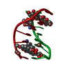
| ||||||||||
| Unit cell |
| ||||||||||
| Details | The second part of the biological assembly is generated by the two fold axis: -Y,-X,1/2-Z |
-Components
| #1: DNA chain | Mass: 1809.217 Da / Num. of mol.: 1 / Source method: obtained synthetically | ||
|---|---|---|---|
| #2: Chemical | ChemComp-DM2 /   Mass: 543.519 Da / Num. of mol.: 1 / Source method: obtained synthetically / Formula: C27H29NO11 / Comment: medication, chemotherapy*YM Mass: 543.519 Da / Num. of mol.: 1 / Source method: obtained synthetically / Formula: C27H29NO11 / Comment: medication, chemotherapy*YM | ||
| #3: Chemical |   Mass: 204.383 Da / Num. of mol.: 3 / Source method: obtained synthetically / Formula: Tl Mass: 204.383 Da / Num. of mol.: 3 / Source method: obtained synthetically / Formula: Tl#4: Water | ChemComp-HOH / |  Mass: 18.015 Da / Num. of mol.: 58 / Source method: isolated from a natural source / Formula: H2O Mass: 18.015 Da / Num. of mol.: 58 / Source method: isolated from a natural source / Formula: H2O |
-Experimental details
-Experiment
| Experiment | Method: X-RAY DIFFRACTION / Number of used crystals: 1 |
|---|
- Sample preparation
| Crystal | Density Matthews: 2.69 Å3/Da / Density % sol: 54.29 % | ||||||||||||||||||||||||||||||||||||||||||||||||
|---|---|---|---|---|---|---|---|---|---|---|---|---|---|---|---|---|---|---|---|---|---|---|---|---|---|---|---|---|---|---|---|---|---|---|---|---|---|---|---|---|---|---|---|---|---|---|---|---|---|
| Crystal grow | Temperature: 295 K / Method: vapor diffusion, hanging drop / pH: 6.4 Details: d(CGATCG), Tl acetate, Mg acetate, MPD, spermine acetate, adriamycin, pH 6.4, VAPOR DIFFUSION, HANGING DROP, temperature 295K | ||||||||||||||||||||||||||||||||||||||||||||||||
| Components of the solutions |
| ||||||||||||||||||||||||||||||||||||||||||||||||
| Crystal grow | *PLUS Method: vapor diffusion, sitting drop | ||||||||||||||||||||||||||||||||||||||||||||||||
| Components of the solutions | *PLUS
|
-Data collection
| Diffraction | Mean temperature: 93 K |
|---|---|
| Diffraction source | Source: ROTATING ANODE / Type: RIGAKU RUH3R / Wavelength: 1.5418 Å |
| Detector | Type: RIGAKU RAXIS IV / Detector: IMAGE PLATE / Date: Aug 3, 2001 / Details: Osmic blue multilayer confocal |
| Radiation | Protocol: SINGLE WAVELENGTH / Monochromatic (M) / Laue (L): M / Scattering type: x-ray |
| Radiation wavelength | Wavelength: 1.5418 Å / Relative weight: 1 |
| Reflection | Resolution: 1.26→100 Å |
| Reflection | *PLUS Lowest resolution: 100 Å / Num. obs: 5559 / % possible obs: 99.9 % / Num. measured all: 31250 |
| Reflection shell | *PLUS Highest resolution: 1.34 Å / % possible obs: 99.9 % |
- Processing
| Software |
| |||||||||||||||||||||||||
|---|---|---|---|---|---|---|---|---|---|---|---|---|---|---|---|---|---|---|---|---|---|---|---|---|---|---|
| Refinement | Method to determine structure: MOLECULAR REPLACEMENT Starting model: NDB Entry DDF044 Resolution: 1.34→35 Å / Isotropic thermal model: isotropic Cross valid method: throughout refinement except final round σ(F): 2 / Stereochemistry target values: Parkinson & Berman, 1996
| |||||||||||||||||||||||||
| Displacement parameters | Biso mean: 13.8 Å2 | |||||||||||||||||||||||||
| Refinement step | Cycle: LAST / Resolution: 1.34→35 Å
| |||||||||||||||||||||||||
| Refine LS restraints |
| |||||||||||||||||||||||||
| Refinement | *PLUS Lowest resolution: 35 Å | |||||||||||||||||||||||||
| Solvent computation | *PLUS | |||||||||||||||||||||||||
| Displacement parameters | *PLUS |
