- EMDB-2316: Structural Basis of Signal Sequence Surveillance and Selection by... -
+
データを開く
IDまたはキーワード:
読み込み中...
-
基本情報
登録情報
データベース: EMDB / ID: EMD-2316
タイトル
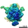
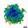
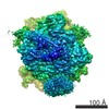
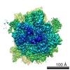
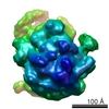
Structural Basis of Signal Sequence Surveillance and Selection by the SRP-SR Complex
マップデータ
RNCEspP-SRP-FtsY
試料
試料: Ribosome-SRP-FtsY complex with EspP nascent chain
複合体: 70S ribosome
タンパク質・ペプチド: SIGNAL RECOGNITION PARTICLE PROTEIN
タンパク質・ペプチド: SIGNAL RECOGNITION PARTICLE RECEPTOR FTSY
タンパク質・ペプチド: 4.5 S RNA
タンパク質・ペプチド: SIGNAL RECOGNITION PARTICLE 54 KDA PROTEIN
タンパク質・ペプチド: DIPEPTIDYL AMINOPEPTIDASE B
キーワード
Ribosome / SRP / signal recognition particle / SR / FtsY / cryo-EM
機能・相同性
機能・相同性情報
Synthesis, secretion, and inactivation of Glucagon-like Peptide-1 (GLP-1) / 加水分解酵素; プロテアーゼ; ペプチド結合加水分解酵素; ジペプチジルペプチターゼ・トリペプチジルペプチターゼ / signal recognition particle / signal recognition particle binding / signal-recognition-particle GTPase / 7S RNA binding / SRP-dependent cotranslational protein targeting to membrane / fungal-type vacuole membrane / dipeptidyl-peptidase activity / stringent response ...Synthesis, secretion, and inactivation of Glucagon-like Peptide-1 (GLP-1) / 加水分解酵素; プロテアーゼ; ペプチド結合加水分解酵素; ジペプチジルペプチターゼ・トリペプチジルペプチターゼ / signal recognition particle / signal recognition particle binding / signal-recognition-particle GTPase / 7S RNA binding / SRP-dependent cotranslational protein targeting to membrane / fungal-type vacuole membrane / dipeptidyl-peptidase activity / stringent response / aminopeptidase activity / protein targeting / protein processing / cytoplasmic side of plasma membrane / serine-type endopeptidase activity / GTPase activity / GTP binding / protein homodimerization activity / ATP hydrolysis activity / proteolysis / plasma membrane / cytosol 類似検索 - 分子機能
Signal-recognition particle receptor FtsY / Signal recognition particle protein / SRP/SRP receptor, N-terminal / Signal recognition particle, SRP54 subunit / Signal recognition particle, SRP54 subunit, M-domain / Signal recognition particle, SRP54 subunit, M-domain superfamily / Signal peptide binding domain / SRP54-type proteins GTP-binding domain signature. / Signal recognition particle SRP54, helical bundle / Signal recognition particle SRP54, N-terminal domain superfamily ...Signal-recognition particle receptor FtsY / Signal recognition particle protein / SRP/SRP receptor, N-terminal / Signal recognition particle, SRP54 subunit / Signal recognition particle, SRP54 subunit, M-domain / Signal recognition particle, SRP54 subunit, M-domain superfamily / Signal peptide binding domain / SRP54-type proteins GTP-binding domain signature. / Signal recognition particle SRP54, helical bundle / Signal recognition particle SRP54, N-terminal domain superfamily / SRP54-type protein, helical bundle domain / SRP54-type protein, helical bundle domain / Signal recognition particle, SRP54 subunit, GTPase domain / SRP54-type protein, GTPase domain / SRP54-type protein, GTPase domain / Prolyl endopeptidase family serine active site. / : / Peptidase S9, serine active site / Dipeptidylpeptidase IV, N-terminal domain / Dipeptidyl peptidase IV (DPP IV) N-terminal region / Peptidase S9, prolyl oligopeptidase, catalytic domain / Prolyl oligopeptidase family / Alpha/Beta hydrolase fold / ATPases associated with a variety of cellular activities / AAA+ ATPase domain / P-loop containing nucleoside triphosphate hydrolase 類似検索 - ドメイン・相同性
Signal recognition particle protein / Signal recognition particle receptor FtsY / Dipeptidyl aminopeptidase B / Signal recognition particle 54 kDa protein 類似検索 - 構成要素
ジャーナル: Nat Struct Mol Biol / 年: 2013 タイトル: Structural basis of signal sequence surveillance and selection by the SRP-FtsY complex. 著者: Ottilie von Loeffelholz / Kèvin Knoops / Aileen Ariosa / Xin Zhang / Manikandan Karuppasamy / Karine Huard / Guy Schoehn / Imre Berger / Shu-ou Shan / Christiane Schaffitzel / 要旨: Signal-recognition particle (SRP)-dependent targeting of translating ribosomes to membranes is a multistep quality-control process. Ribosomes that are translating weakly hydrophobic signal sequences ...Signal-recognition particle (SRP)-dependent targeting of translating ribosomes to membranes is a multistep quality-control process. Ribosomes that are translating weakly hydrophobic signal sequences can be rejected from the targeting reaction even after they are bound to the SRP. Here we show that the early complex, formed by Escherichia coli SRP and its receptor FtsY with ribosomes translating the incorrect cargo EspP, is unstable and rearranges inefficiently into subsequent conformational states, such that FtsY dissociation is favored over successful targeting. The N-terminal extension of EspP is responsible for these defects in the early targeting complex. The cryo-electron microscopy structure of this 'false' early complex with EspP revealed an ordered M domain of SRP protein Ffh making two ribosomal contacts, and the NG domains of Ffh and FtsY forming a distorted, flexible heterodimer. Our results provide a structural basis for SRP-mediated signal-sequence selection during recruitment of the SRP receptor.
pH: 7.5 / 詳細: 50 mM Hepes-KOH, 100 mM KOAc, 8 mM Mg(OAc)2, pH 7.5
凍結
凍結剤: NITROGEN / チャンバー内湿度: 100 % / チャンバー内温度: 77 K / 装置: FEI VITROBOT MARK IV 手法: grids were glow discharged on both sides for 30 s, blottime 1s, blotforce 1
-
電子顕微鏡法
顕微鏡
FEI POLARA 300
温度
最低: 77 K / 最高: 80 K / 平均: 78 K
アライメント法
Legacy - 非点収差: correction based on power spectrum from images taken at 100000 x magnification
詳細
low dose
日付
2011年1月11日
撮影
カテゴリ: CCD フィルム・検出器のモデル: GENERIC GATAN (4k x 4k) デジタル化 - サンプリング間隔: 15 µm / 実像数: 1974 / 平均電子線量: 15 e/Å2 / カメラ長: 61.44 / 詳細: Images recorded on CCD camera / ビット/ピクセル: 32
 ムービー
ムービー コントローラー


 データを開く
 基本情報
基本情報 マップデータ
 試料
試料 キーワード
キーワード 機能・相同性情報

 Sulfolobus solfataricus (古細菌) /
 データ登録者
 引用
引用
 構造の表示
構造の表示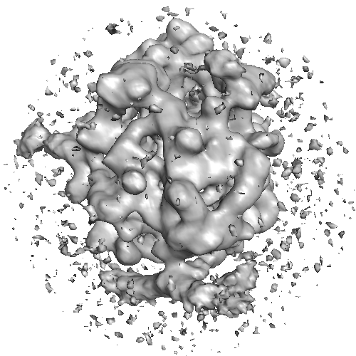
 ダウンロードとリンク
 2316-view_2.png
 http://ftp.pdbj.org/pub/emdb/structures/EMD-2316


 Z (Sec.)
 Y (Row.)
 X (Col.)


 試料の構成要素
 解析
解析 電子顕微鏡法
 FIELD EMISSION GUN
