 ムービー
ムービー コントローラー


+ データを開く

- 基本情報
基本情報
| 登録情報 | データベース: EMDB / ID: EMD-20632 | |||||||||
|---|---|---|---|---|---|---|---|---|---|---|
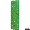
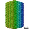
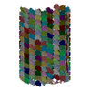
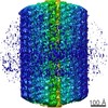
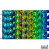
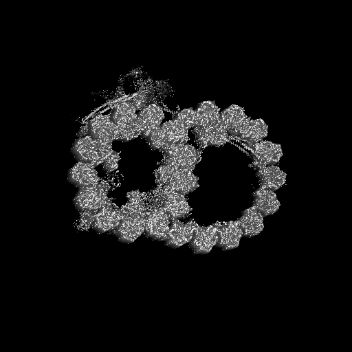
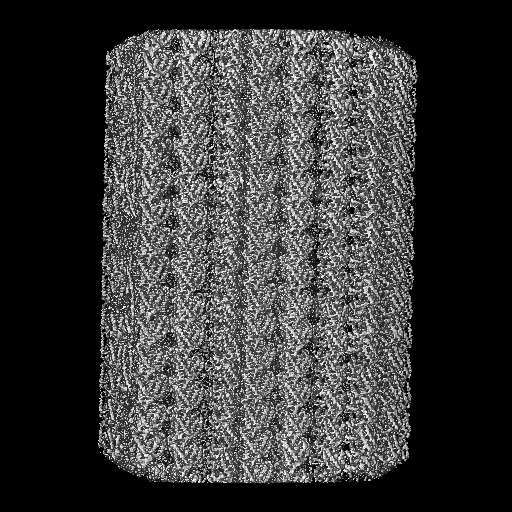
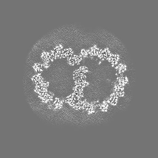
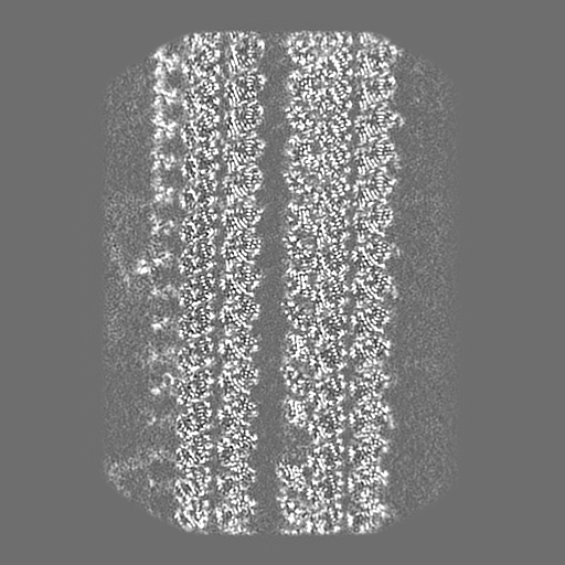
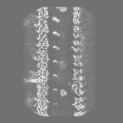
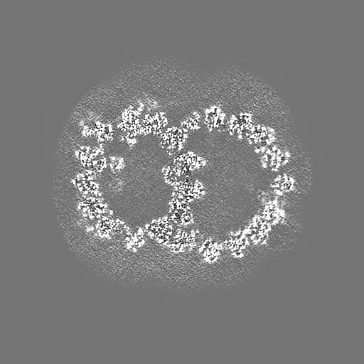
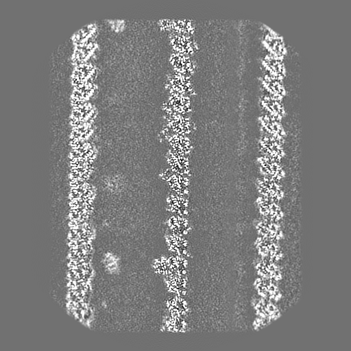
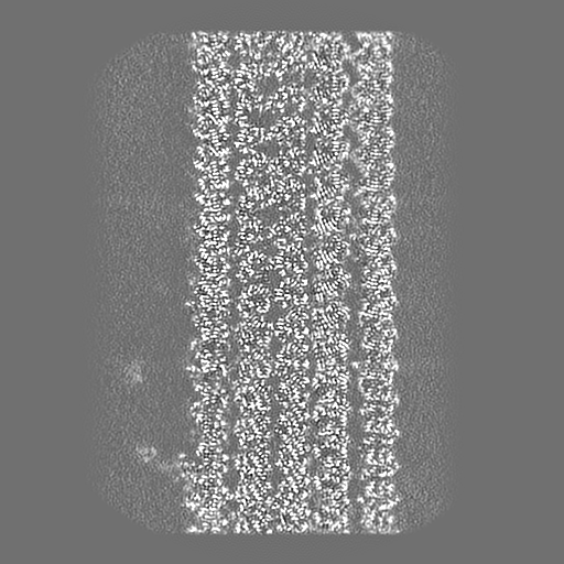
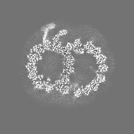
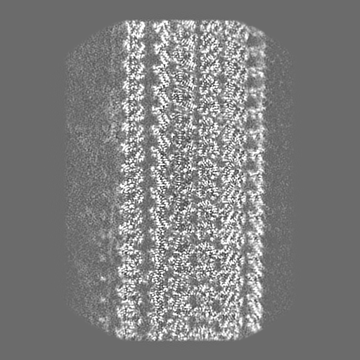
| タイトル | Composite cryo-EM density map of half 1 of the 96-nm repeat of the doublet microtubule from wild-type Chlamydomonas reinhardtii | |||||||||
 マップデータ マップデータ | Composite cryo-EM density map of half 1 of the 96-nm repeat of the doublet microtubule from wild-type Chlamydomonas reinhardtii | |||||||||
 試料 試料 |
| |||||||||
| 生物種 |   Chlamydomonas reinhardtii (クラミドモナス) Chlamydomonas reinhardtii (クラミドモナス) | |||||||||
| 手法 | 単粒子再構成法 / クライオ電子顕微鏡法 / 解像度: 3.6 Å | |||||||||
 データ登録者 データ登録者 | Ma M / Stoyanova M / Rademacher G / Dutcher SK / Brown A / Zhang R | |||||||||
| 資金援助 |  米国, 1件 米国, 1件
| |||||||||
 引用 引用 | ジャーナル: Cell / 年: 2019 タイトル: Structure of the Decorated Ciliary Doublet Microtubule. 著者: Meisheng Ma / Mihaela Stoyanova / Griffin Rademacher / Susan K Dutcher / Alan Brown / Rui Zhang / 要旨: The axoneme of motile cilia is the largest macromolecular machine of eukaryotic cells. In humans, impaired axoneme function causes a range of ciliopathies. Axoneme assembly, structure, and motility ...The axoneme of motile cilia is the largest macromolecular machine of eukaryotic cells. In humans, impaired axoneme function causes a range of ciliopathies. Axoneme assembly, structure, and motility require a radially arranged set of doublet microtubules, each decorated in repeating patterns with non-tubulin components. We use single-particle cryo-electron microscopy to visualize and build an atomic model of the repeating structure of a native axonemal doublet microtubule, which reveals the identities, positions, repeat lengths, and interactions of 38 associated proteins, including 33 microtubule inner proteins (MIPs). The structure demonstrates how these proteins establish the unique architecture of doublet microtubules, maintain coherent periodicities along the axoneme, and stabilize the microtubules against the repeated mechanical stress induced by ciliary motility. Our work elucidates the architectural principles that underpin the assembly of this large, repetitive eukaryotic structure and provides a molecular basis for understanding the etiology of human ciliopathies. | |||||||||
| 履歴 |
|
- 構造の表示
構造の表示
| ムービー |
 ムービービューア ムービービューア |
|---|---|
| 構造ビューア | EMマップ: SurfViewMolmilJmol/JSmol |
| 添付画像 |

- ダウンロードとリンク
-EMDBアーカイブ
| マップデータ | emd_20632.map.gz | 203.9 MB | EMDBマップデータ形式 | |
|---|---|---|---|---|
| ヘッダ (付随情報) | emd-20632-v30.xmlemd-20632.xml | 17.7 KB 17.7 KB | 表示 表示 | EMDBヘッダ |
| FSC (解像度算出) | emd_20632_fsc.xml | 18 KB | 表示 | FSCデータファイル |
| 画像 | 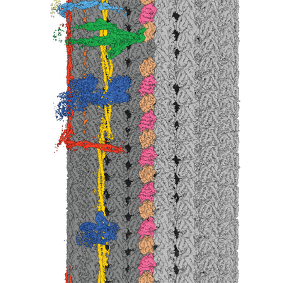 emd_20632.png emd_20632.png | 243 KB | ||
| マスクデータ | emd_20632_msk_1.map | 512 MB | マスクマップ | |
| その他 | emd_20632_half_map_1.map.gzemd_20632_half_map_2.map.gz | 175.1 MB 175.3 MB | ||
| アーカイブディレクトリ |  http://ftp.pdbj.org/pub/emdb/structures/EMD-20632ftp://ftp.pdbj.org/pub/emdb/structures/EMD-20632 http://ftp.pdbj.org/pub/emdb/structures/EMD-20632ftp://ftp.pdbj.org/pub/emdb/structures/EMD-20632 | HTTPS FTP |
-検証レポート
| 文書・要旨 | emd_20632_validation.pdf.gz | 78.5 KB | 表示 | EMDB検証レポート |
|---|---|---|---|---|
| 文書・詳細版 | emd_20632_full_validation.pdf.gz | 77.6 KB | 表示 | |
| XML形式データ | emd_20632_validation.xml.gz | 494 B | 表示 | |
| アーカイブディレクトリ | https://ftp.pdbj.org/pub/emdb/validation_reports/EMD-20632ftp://ftp.pdbj.org/pub/emdb/validation_reports/EMD-20632 | HTTPS FTP |
-関連構造データ
-リンク
| EMDBのページ | EMDB (EBI/PDBe) / EMDataResource |
|---|
-マップ
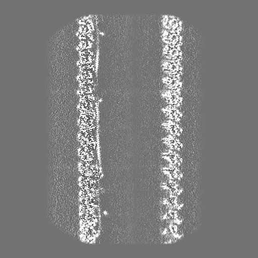
| ファイル | ダウンロード / ファイル: emd_20632.map.gz / 形式: CCP4 / 大きさ: 512 MB / タイプ: IMAGE STORED AS FLOATING POINT NUMBER (4 BYTES) | ||||||||||||||||||||||||||||||||||||||||||||||||||||||||||||
|---|---|---|---|---|---|---|---|---|---|---|---|---|---|---|---|---|---|---|---|---|---|---|---|---|---|---|---|---|---|---|---|---|---|---|---|---|---|---|---|---|---|---|---|---|---|---|---|---|---|---|---|---|---|---|---|---|---|---|---|---|---|
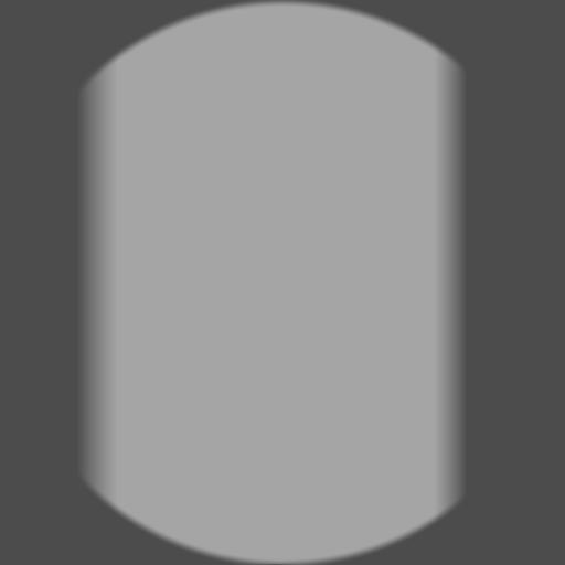
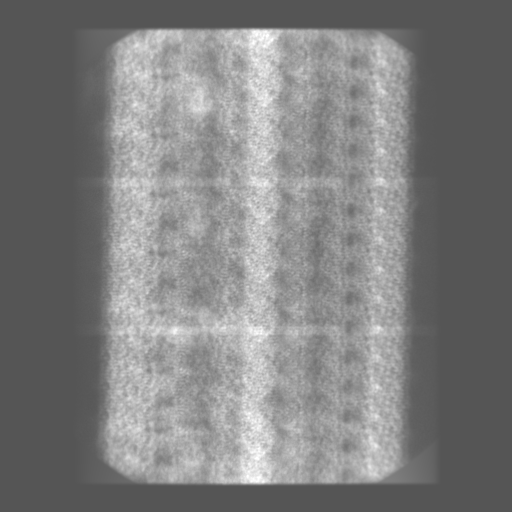
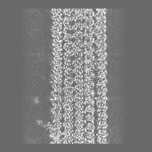
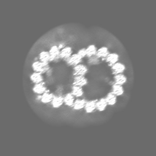
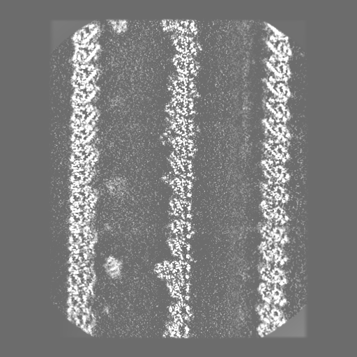
| 注釈 | Composite cryo-EM density map of half 1 of the 96-nm repeat of the doublet microtubule from wild-type Chlamydomonas reinhardtii | ||||||||||||||||||||||||||||||||||||||||||||||||||||||||||||
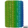
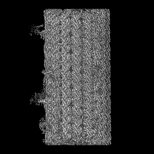
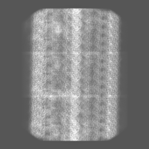
| 投影像・断面図 | 画像のコントロール
画像は Spider により作成 | ||||||||||||||||||||||||||||||||||||||||||||||||||||||||||||
| ボクセルのサイズ | X=Y=Z: 1.403 Å | ||||||||||||||||||||||||||||||||||||||||||||||||||||||||||||
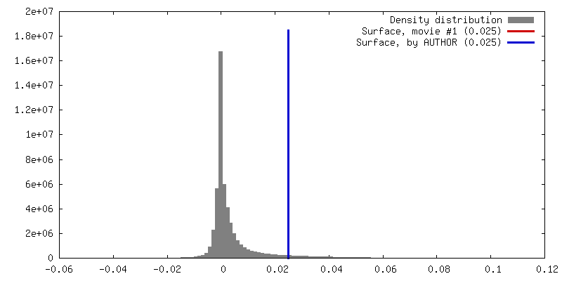
| 密度 |
| ||||||||||||||||||||||||||||||||||||||||||||||||||||||||||||
| 対称性 | 空間群: 1 | ||||||||||||||||||||||||||||||||||||||||||||||||||||||||||||
| 詳細 | EMDB XML:
CCP4マップ ヘッダ情報:
| ||||||||||||||||||||||||||||||||||||||||||||||||||||||||||||
 Z (Sec.)
 Y (Row.)
 X (Col.)


-添付データ
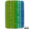
-マスク #1
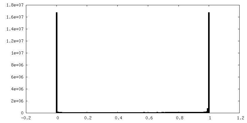
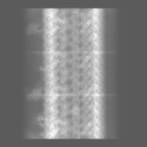
| ファイル | emd_20632_msk_1.map | ||||||||||||
|---|---|---|---|---|---|---|---|---|---|---|---|---|---|
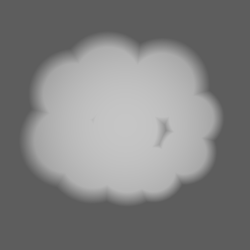
| 投影像・断面図 |
| ||||||||||||
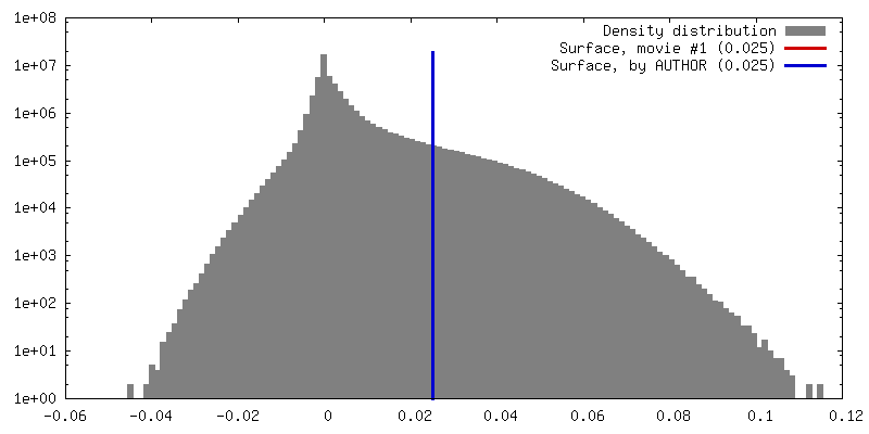
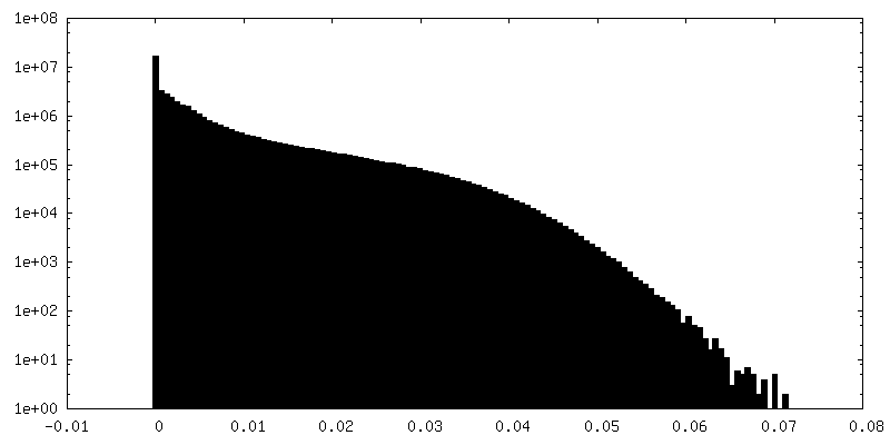
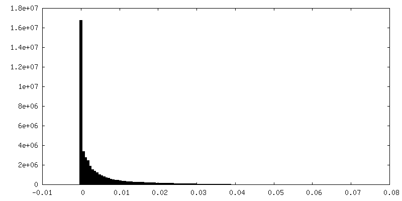
| 密度ヒストグラム |


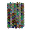
-ハーフマップ: half map 1 of the composite cryo-EM density...
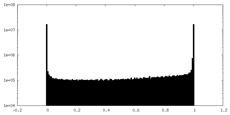
| ファイル | emd_20632_half_map_1.map | ||||||||||||
|---|---|---|---|---|---|---|---|---|---|---|---|---|---|
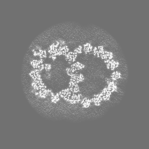
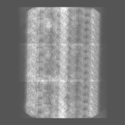
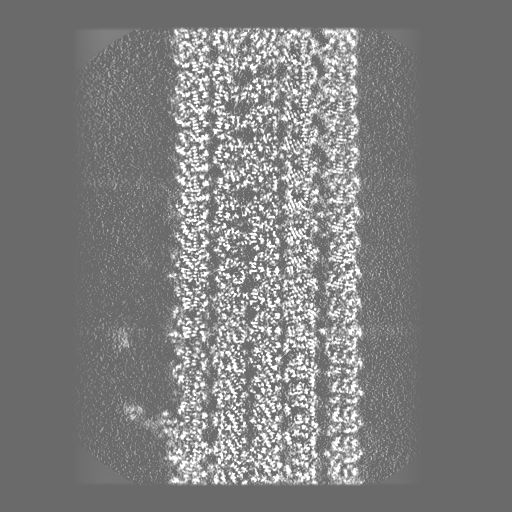
| 注釈 | half map 1 of the composite cryo-EM density map of half 1 of the 96-nm repeat of the doublet microtubule from wild-type Chlamydomonas reinhardtii | ||||||||||||
| 投影像・断面図 |
| ||||||||||||
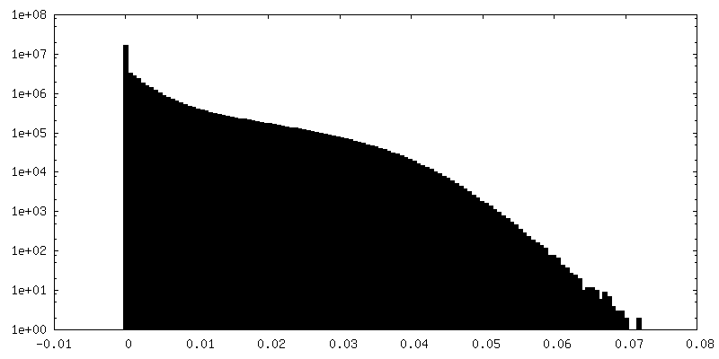
| 密度ヒストグラム |

-ハーフマップ: half map 1 of the composite cryo-EM density...
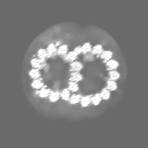
| ファイル | emd_20632_half_map_2.map | ||||||||||||
|---|---|---|---|---|---|---|---|---|---|---|---|---|---|
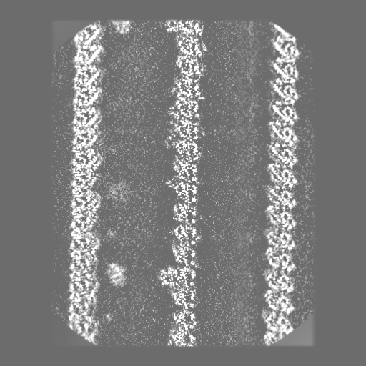
| 注釈 | half map 1 of the composite cryo-EM density map of half 1 of the 96-nm repeat of the doublet microtubule from wild-type Chlamydomonas reinhardtii | ||||||||||||
| 投影像・断面図 |
| ||||||||||||
| 密度ヒストグラム |


- 試料の構成要素
-全体 : doublet microtubule isolated from wild-type Chlamydomonas reinhardtii
| 全体 | 名称: doublet microtubule isolated from wild-type Chlamydomonas reinhardtii |
|---|---|
| 要素 |
|
-超分子 #1: doublet microtubule isolated from wild-type Chlamydomonas reinhardtii
| 超分子 | 名称: doublet microtubule isolated from wild-type Chlamydomonas reinhardtii タイプ: complex / ID: 1 / 親要素: 0 |
|---|---|
| 由来(天然) | 生物種: Chlamydomonas reinhardtii (クラミドモナス) 株: CC-125 |
-実験情報
-構造解析
| 手法 | クライオ電子顕微鏡法 |
|---|---|
 解析 解析 | 単粒子再構成法 |
| 試料の集合状態 | filament |
-試料調製
| 緩衝液 | pH: 7.4 / 構成要素 - 名称: HMDEKP 詳細: 30 mM HEPES, 5 mM MgSO4, 1 mM DTT, 0.5 mM EGTA, 25 mM KCl, PH 7.4 |
|---|---|
| グリッド | モデル: C-flat-1.2/1.3 4C / 材質: COPPER / メッシュ: 400 / 前処理 - タイプ: GLOW DISCHARGE / 前処理 - 雰囲気: AIR / 詳細: The grid was glow discharged using PELCO easiGlow |
| 凍結 | 凍結剤: ETHANE / チャンバー内湿度: 95 % / チャンバー内温度: 277 K / 装置: FEI VITROBOT MARK IV / 詳細: blot for 4 seconds before plunging. |
- 電子顕微鏡法
| 顕微鏡 | FEI TITAN KRIOS |
|---|---|
| 特殊光学系 | 球面収差補正装置: Microscope is equipped with a Cs corrector エネルギーフィルター - 名称: GIF Bioquantum / エネルギーフィルター - スリット幅: 20 eV |
| 撮影 | フィルム・検出器のモデル: GATAN K2 QUANTUM (4k x 4k) 検出モード: COUNTING / デジタル化 - サイズ - 横: 3838 pixel / デジタル化 - サイズ - 縦: 3710 pixel / デジタル化 - 画像ごとのフレーム数: 1-30 / 撮影したグリッド数: 6 / 実像数: 8314 / 平均露光時間: 9.0 sec. / 平均電子線量: 38.9 e/Å2 |
| 電子線 | 加速電圧: 300 kV / 電子線源:  FIELD EMISSION GUN FIELD EMISSION GUN |
| 電子光学系 | 最大 デフォーカス(補正後): 3.1 µm / 最小 デフォーカス(補正後): 1.0 µm / 倍率(補正後): 81000 / 照射モード: FLOOD BEAM / 撮影モード: BRIGHT FIELD / Cs: 0.01 mm / 最大 デフォーカス(公称値): 2.75 µm / 最小 デフォーカス(公称値): 1.25 µm / 倍率(公称値): 81000 |
| 試料ステージ | 試料ホルダーモデル: FEI TITAN KRIOS AUTOGRID HOLDER ホルダー冷却材: NITROGEN |
| 実験機器 |  モデル: Titan Krios / 画像提供: FEI Company |
+画像解析

-原子モデル構築 1
| 精密化 | 空間: REAL / プロトコル: AB INITIO MODEL / 温度因子: 50 |
|---|
