 Movie
Movie Controller

[English] 日本語
 Yorodumi
- PDB-1vaf: Inducible nitric oxide synthase oxygenase domain complexed with t... -
+ Open data

- Basic information
| Entry | Database: PDB / ID: 1vaf | ||||||
|---|---|---|---|---|---|---|---|
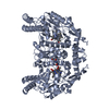
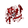
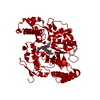
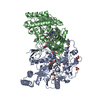
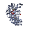
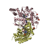
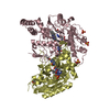
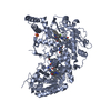
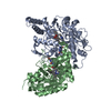
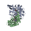
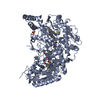
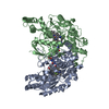
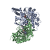
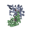
| Title | Inducible nitric oxide synthase oxygenase domain complexed with the inhibitor AR-R17477 | ||||||
 Components Components | Nitric oxide synthase, inducible | ||||||
 Keywords Keywords | OXIDOREDUCTASE / MURINE INOSOXY INHIBITOR COMPLEX | ||||||
| Function / homology |  Function and homology information Function and homology informationNitric oxide stimulates guanylate cyclase / ROS and RNS production in phagocytes / peptidyl-cysteine S-nitrosylation / Peroxisomal protein import / prostaglandin secretion / tetrahydrobiopterin binding / arginine binding / superoxide metabolic process / regulation of cytokine production involved in inflammatory response / cellular response to cytokine stimulus ...Nitric oxide stimulates guanylate cyclase / ROS and RNS production in phagocytes / peptidyl-cysteine S-nitrosylation / Peroxisomal protein import / prostaglandin secretion / tetrahydrobiopterin binding / arginine binding / superoxide metabolic process / regulation of cytokine production involved in inflammatory response / cellular response to cytokine stimulus / Fc-gamma receptor signaling pathway involved in phagocytosis / cortical cytoskeleton / nitric-oxide synthase (NADPH) / nitric-oxide synthase activity / L-arginine catabolic process / nitric oxide biosynthetic process / regulation of insulin secretion / positive regulation of interleukin-8 production / response to bacterium / circadian rhythm / negative regulation of protein catabolic process / cellular response to type II interferon / positive regulation of interleukin-6 production / cellular response to xenobiotic stimulus / peroxisome / FMN binding / NADP binding / flavin adenine dinucleotide binding / regulation of cell population proliferation / cellular response to lipopolysaccharide / response to lipopolysaccharide / response to hypoxia / calmodulin binding / defense response to bacterium / inflammatory response / negative regulation of gene expression / heme binding / perinuclear region of cytoplasm / protein homodimerization activity / metal ion binding / cytosol / cytoplasm Similarity search - Function | ||||||
| Biological species |  | ||||||
| Method |  X-RAY DIFFRACTION / SYNCHROTRON / MOLECULAR REPLACEMENT / Resolution: 2.9 Å X-RAY DIFFRACTION / SYNCHROTRON / MOLECULAR REPLACEMENT / Resolution: 2.9 Å | ||||||
 Authors Authors | Fedorov, R. / Vasan, R. / Ghosh, D.K. / Schlichting, I. | ||||||
 Citation Citation | Journal: Proc.Natl.Acad.Sci.USA / Year: 2004 Title: Structures of nitric oxide synthase isoforms complexed with the inhibitor AR-R17477 suggest a rational basis for specificity and inhibitor design Authors: Fedorov, R. / Vasan, R. / Ghosh, D.K. / Schlichting, I. | ||||||
| History |
|
- Structure visualization
| Structure viewer | Molecule: MolmilJmol/JSmol |
|---|
- Downloads & links
-Download
| PDBx/mmCIF format | 1vaf.cif.gz | 190.6 KB | Display | PDBx/mmCIF format |
|---|---|---|---|---|
| PDB format | pdb1vaf.ent.gz | 151.8 KB | Display | PDB format |
| PDBx/mmJSON format | 1vaf.json.gz | Tree view | PDBx/mmJSON format | |
| Others |  Other downloads Other downloads |
-Validation report
| Arichive directory | https://data.pdbj.org/pub/pdb/validation_reports/va/1vafftp://data.pdbj.org/pub/pdb/validation_reports/va/1vaf | HTTPS FTP |
|---|
-Related structure data
| Related structure data | 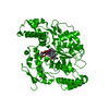 1vagC 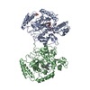 1nodS C: citing same article ( S: Starting model for refinement |
|---|---|
| Similar structure data |


-Links
 PDBj
PDBj


- Assembly
| Deposited unit | 
| ||||||||
|---|---|---|---|---|---|---|---|---|---|
| 1 | 
| ||||||||
| 2 | 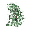
| ||||||||
| Unit cell |
| ||||||||
| Components on special symmetry positions |
| ||||||||
| Details | The dimer in asymmetric unit is not a biological dimer, but the counterpart of the biological dimer for molecule having chain ID "A" can be obtained by application of the symmetry operation: (-y, -x, -z+5/6 ) and for molecule with chain ID "B": (-x, -x+y, -z+2/3 ). |
-Components
-Protein , 1 types, 2 molecules AB
| #1: Protein | Mass: 48498.168 Da / Num. of mol.: 2 Fragment: Inducible Nitric Oxide Synthase Oxygenase Domain (residues 77-495) Source method: isolated from a genetically manipulated source Source: (gene. exp.)  |
|---|
-Non-polymers , 5 types, 189 molecules 


| #2: Chemical |  Mass: 65.409 Da / Num. of mol.: 2 / Source method: obtained synthetically / Formula: Zn Mass: 65.409 Da / Num. of mol.: 2 / Source method: obtained synthetically / Formula: Zn#3: Chemical |  Mass: 616.487 Da / Num. of mol.: 2 / Source method: obtained synthetically / Formula: C34H32FeN4O4 Mass: 616.487 Da / Num. of mol.: 2 / Source method: obtained synthetically / Formula: C34H32FeN4O4#4: Chemical |  Mass: 241.247 Da / Num. of mol.: 2 / Source method: obtained synthetically / Formula: C9H15N5O3 / Comment: neurotransmitter*YM Mass: 241.247 Da / Num. of mol.: 2 / Source method: obtained synthetically / Formula: C9H15N5O3 / Comment: neurotransmitter*YM#5: Chemical |  Mass: 369.911 Da / Num. of mol.: 2 / Source method: obtained synthetically / Formula: C20H20ClN3S Mass: 369.911 Da / Num. of mol.: 2 / Source method: obtained synthetically / Formula: C20H20ClN3S#6: Water | ChemComp-HOH / | Mass: 18.015 Da / Num. of mol.: 181 / Source method: isolated from a natural source / Formula: H2O |
|---|
-Experimental details
-Experiment
| Experiment | Method: X-RAY DIFFRACTION / Number of used crystals: 1 |
|---|
- Sample preparation
| Crystal | Density Matthews: 3.93 Å3/Da / Density % sol: 68.46 % |
|---|---|
| Crystal grow | Temperature: 277 K / Method: vapor diffusion, hanging drop / pH: 7 Details: MES, SODIUM-MALONATE, EPPS, NACL, GLYCEROL, H4B, beta-MERCAPTOETHANOL, AR-R17477, pH 7.0, VAPOR DIFFUSION, HANGING DROP, temperature 277K |
-Data collection
| Diffraction | Mean temperature: 100 K |
|---|---|
| Diffraction source | Source: SYNCHROTRON / Site: ESRF  / Beamline: ID14-1 / Wavelength: 0.934 Å / Beamline: ID14-1 / Wavelength: 0.934 Å |
| Detector | Type: ADSC QUANTUM 4 / Detector: CCD / Date: Sep 1, 2001 |
| Radiation | Protocol: SINGLE WAVELENGTH / Monochromatic (M) / Laue (L): M / Scattering type: x-ray |
| Radiation wavelength | Wavelength: 0.934 Å / Relative weight: 1 |
| Reflection | Resolution: 2.9→20 Å / Num. all: 33741 / Num. obs: 33439 / % possible obs: 96.5 % / Observed criterion σ(F): 2 / Observed criterion σ(I): 2 / Redundancy: 7.3 % / Rsym value: 0.058 / Net I/σ(I): 21.6 |
| Reflection shell | Resolution: 2.9→3 Å / Mean I/σ(I) obs: 5.7 / Rsym value: 0.381 / % possible all: 95.8 |
- Processing
| Software |
| ||||||||||||||||||||
|---|---|---|---|---|---|---|---|---|---|---|---|---|---|---|---|---|---|---|---|---|---|
| Refinement | Method to determine structure: MOLECULAR REPLACEMENT Starting model: PDB entry 1NOD Resolution: 2.9→8 Å / Cross valid method: THROUGHOUT / σ(F): 0 / Stereochemistry target values: Engh & Huber
| ||||||||||||||||||||
| Refinement step | Cycle: LAST / Resolution: 2.9→8 Å
|
