 Movie
Movie Controller

[English] 日本語
 Yorodumi
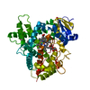
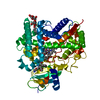
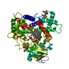
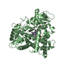
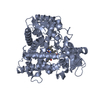
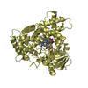
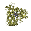
- PDB-1suo: Structure of mammalian cytochrome P450 2B4 with bound 4-(4-chloro... -
+ Open data

- Basic information
| Entry | Database: PDB / ID: 1suo | ||||||
|---|---|---|---|---|---|---|---|
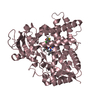
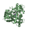
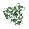
| Title | Structure of mammalian cytochrome P450 2B4 with bound 4-(4-chlorophenyl)imidazole | ||||||
 Components Components | Cytochrome P450 2B4 | ||||||
 Keywords Keywords | OXIDOREDUCTASE / membrane protein / CYP 2B4 / CYP LM2 / Cytochrome p450 / Monooxygenase | ||||||
| Function / homology |  Function and homology information Function and homology informationarachidonate epoxygenase activity / epoxygenase P450 pathway / oxidoreductase activity, acting on paired donors, with incorporation or reduction of molecular oxygen, reduced flavin or flavoprotein as one donor, and incorporation of one atom of oxygen / unspecific monooxygenase / xenobiotic metabolic process / iron ion binding / heme binding / endoplasmic reticulum membrane Similarity search - Function | ||||||
| Biological species |  | ||||||
| Method |  X-RAY DIFFRACTION / SYNCHROTRON / MOLECULAR REPLACEMENT / Resolution: 1.9 Å X-RAY DIFFRACTION / SYNCHROTRON / MOLECULAR REPLACEMENT / Resolution: 1.9 Å | ||||||
 Authors Authors | Scott, E.E. / White, M.A. / He, Y.A. / Johnson, E.F. / Stout, C.D. / Halpert, J.R. | ||||||
 Citation Citation | Journal: J.Biol.Chem. / Year: 2004 Title: Structure of mammalian cytochrome P450 2B4 complexed with 4-(4-chlorophenyl)imidazole at 1.9 {angstrom} resolution: Insight into the range of P450 conformations and coordination of redox partner binding. Authors: Scott, E.E. / White, M.A. / He, Y.A. / Johnson, E.F. / Stout, C.D. / Halpert, J.R. #1: Journal: Proc.Natl.Acad.Sci.USA / Year: 2003Title: An Open Conformation of Mammalian Cytochrome P450 2B4 at 1.6 A Resolution Authors: Scott, E.E. / He, Y.A. / Wester, M.R. / White, M.A. / Chin, C.C. / Halpert, J.R. / Johnson, E.F. / Stout, C.D. #2: Journal: Arch.Biochem.Biophys. / Year: 2001Title: A Truncation of 2B Subfamily Cytochromes P450 Yields Increased Expression Levels, Increased Solubility, and Decreased Aggregation While Retaining Function Authors: Scott, E.E. / Spatzenegger, M. / Halpert, J.R. | ||||||
| History |
| ||||||
| Remark 999 | SEQUENCE AUTHORS INFORMED THAT THE GENBANK SEQUENCE IS THOUGHT TO CONTAIN A SEQUENCING ERROR AT RESIDUE 221. |
- Structure visualization
| Structure viewer | Molecule: MolmilJmol/JSmol |
|---|
- Downloads & links
-Download
| PDBx/mmCIF format | 1suo.cif.gz | 115 KB | Display | PDBx/mmCIF format |
|---|---|---|---|---|
| PDB format | pdb1suo.ent.gz | 86.4 KB | Display | PDB format |
| PDBx/mmJSON format | 1suo.json.gz | Tree view | PDBx/mmJSON format | |
| Others |  Other downloads Other downloads |
-Validation report
| Summary document | 1suo_validation.pdf.gz | 821.1 KB | Display | wwPDB validaton report |
|---|---|---|---|---|
| Full document | 1suo_full_validation.pdf.gz | 828 KB | Display | |
| Data in XML | 1suo_validation.xml.gz | 20.8 KB | Display | |
| Data in CIF | 1suo_validation.cif.gz | 30 KB | Display | |
| Arichive directory | https://data.pdbj.org/pub/pdb/validation_reports/su/1suoftp://data.pdbj.org/pub/pdb/validation_reports/su/1suo | HTTPS FTP |
-Related structure data
| Related structure data | 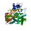 1po5S S: Starting model for refinement |
|---|---|
| Similar structure data |


-Links
 PDBj
PDBj


- Assembly
| Deposited unit | 
| ||||||||
|---|---|---|---|---|---|---|---|---|---|
| 1 |
| ||||||||
| Unit cell |
| ||||||||
| Components on special symmetry positions |
|
-Components
| #1: Protein | Mass: 54169.082 Da / Num. of mol.: 1 Mutation: E2A, G22K, H23K, P24T, K25S, A26S, H27K, R29K, P221S, H226Y Source method: isolated from a genetically manipulated source Details: CYS 436 BINDS HEME IRON. 4-(4-CHLOROPHENYL)IMIDAZOLE IS BOUND IN THE ACTIVE SITE COORDINATING TO THE HEME IRON AS THE SIXTH LIGAND. Source: (gene. exp.)  |
|---|---|
| #2: Chemical | ChemComp-HEM /   Mass: 616.487 Da / Num. of mol.: 1 / Source method: obtained synthetically / Formula: C34H32FeN4O4 Mass: 616.487 Da / Num. of mol.: 1 / Source method: obtained synthetically / Formula: C34H32FeN4O4 |
| #3: Chemical | ChemComp-CPZ / 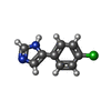  Mass: 178.618 Da / Num. of mol.: 1 / Source method: obtained synthetically / Formula: C9H7ClN2 Mass: 178.618 Da / Num. of mol.: 1 / Source method: obtained synthetically / Formula: C9H7ClN2 |
| #4: Water | ChemComp-HOH /  Mass: 18.015 Da / Num. of mol.: 190 / Source method: isolated from a natural source / Formula: H2O Mass: 18.015 Da / Num. of mol.: 190 / Source method: isolated from a natural source / Formula: H2O |
-Experimental details
-Experiment
| Experiment | Method: X-RAY DIFFRACTION / Number of used crystals: 1 |
|---|
- Sample preparation
| Crystal | Density Matthews: 4.09 Å3/Da / Density % sol: 69.92 % |
|---|---|
| Crystal grow | Temperature: 291 K / Method: vapor diffusion, hanging drop / pH: 4 Details: NACL, PEG 10,000, PHOSPHATE-CITRATE PH 4.0, GLYCEROL, VAPOR DIFFUSION, HANGING DROP, TEMPERATURE 291K |
-Data collection
| Diffraction | Mean temperature: 100 K |
|---|---|
| Diffraction source | Source: SYNCHROTRON / Site: ALS  / Beamline: 5.0.2 / Wavelength: 1.0001 Å / Beamline: 5.0.2 / Wavelength: 1.0001 Å |
| Detector | Type: ADSC QUANTUM 210 / Detector: CCD / Date: Oct 11, 2003 |
| Radiation | Protocol: SINGLE WAVELENGTH / Monochromatic (M) / Laue (L): M / Scattering type: x-ray |
| Radiation wavelength | Wavelength: 1.0001 Å / Relative weight: 1 |
| Reflection | Resolution: 1.9→49 Å / Num. obs: 638636 / % possible obs: 99.9 % / Redundancy: 8.9 % / Rmerge(I) obs: 0.074 / Rsym value: 0.074 / Net I/σ(I): 6.8 |
| Reflection shell | Resolution: 1.9→1.95 Å / Redundancy: 6 % / Rmerge(I) obs: 0.553 / Mean I/σ(I) obs: 1.3 / % possible all: 100 |
- Processing
| Software |
| |||||||||||||||||||||||||
|---|---|---|---|---|---|---|---|---|---|---|---|---|---|---|---|---|---|---|---|---|---|---|---|---|---|---|
| Refinement | Method to determine structure: MOLECULAR REPLACEMENT Starting model: PDB ENTRY 1PO5 Resolution: 1.9→49 Å / Cross valid method: THROUGHOUT / Stereochemistry target values: Engh & Huber
| |||||||||||||||||||||||||
| Refinement step | Cycle: LAST / Resolution: 1.9→49 Å
| |||||||||||||||||||||||||
| Refine LS restraints |
| |||||||||||||||||||||||||
| LS refinement shell | Resolution: 1.9→1.95 Å / % reflection obs: 100 % |
