 ムービー
ムービー コントローラー


+ データを開く

- 基本情報
基本情報
| 登録情報 | データベース: PDB / ID: 1opk | ||||||
|---|---|---|---|---|---|---|---|
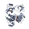
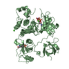
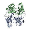
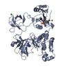
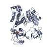
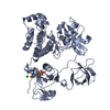
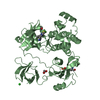
| タイトル | Structural basis for the auto-inhibition of c-Abl tyrosine kinase | ||||||
 要素 要素 | Proto-oncogene tyrosine-protein kinase ABL1 | ||||||
 キーワード キーワード | TRANSFERASE | ||||||
| 機能・相同性 |  機能・相同性情報 機能・相同性情報Role of ABL in ROBO-SLIT signaling / HDR through Single Strand Annealing (SSA) / RHO GTPases Activate WASPs and WAVEs / Cyclin D associated events in G1 / MLL4 and MLL3 complexes regulate expression of PPARG target genes in adipogenesis and hepatic steatosis / Recruitment and ATM-mediated phosphorylation of repair and signaling proteins at DNA double strand breaks / Turbulent (oscillatory, disturbed) flow shear stress activates signaling by PIEZO1 and integrins in endothelial cells / protein localization to cytoplasmic microtubule plus-end / DNA conformation change / DN4 thymocyte differentiation ...Role of ABL in ROBO-SLIT signaling / HDR through Single Strand Annealing (SSA) / RHO GTPases Activate WASPs and WAVEs / Cyclin D associated events in G1 / MLL4 and MLL3 complexes regulate expression of PPARG target genes in adipogenesis and hepatic steatosis / Recruitment and ATM-mediated phosphorylation of repair and signaling proteins at DNA double strand breaks / Turbulent (oscillatory, disturbed) flow shear stress activates signaling by PIEZO1 and integrins in endothelial cells / protein localization to cytoplasmic microtubule plus-end / DNA conformation change / DN4 thymocyte differentiation / RUNX1 regulates transcription of genes involved in differentiation of HSCs / response to epinephrine / phospholipase C-inhibiting G protein-coupled receptor signaling pathway / podocyte apoptotic process / delta-catenin binding / transitional one stage B cell differentiation / regulation of cellular senescence / regulation of postsynaptic specialization assembly / regulation of modification of synaptic structure / cerebellum morphogenesis / neuroepithelial cell differentiation / B cell proliferation involved in immune response / positive regulation of extracellular matrix organization / positive regulation of Wnt signaling pathway, planar cell polarity pathway / Regulation of actin dynamics for phagocytic cup formation / microspike assembly / B-1 B cell homeostasis / neuropilin signaling pathway / neuropilin binding / regulation of extracellular matrix organization / Myogenesis / bubble DNA binding / positive regulation of establishment of T cell polarity / activated T cell proliferation / positive regulation of blood vessel branching / proline-rich region binding / circulatory system development / negative regulation of mitotic cell cycle / regulation of Cdc42 protein signal transduction / mitogen-activated protein kinase binding / syntaxin binding / alpha-beta T cell differentiation / positive regulation of dendrite development / positive regulation of cell migration involved in sprouting angiogenesis / regulation of axon extension / regulation of T cell differentiation / positive regulation of peptidyl-tyrosine phosphorylation / negative regulation of cell-cell adhesion / neuromuscular process controlling balance / positive regulation of osteoblast proliferation / platelet-derived growth factor receptor-beta signaling pathway / positive regulation of vasoconstriction / platelet-derived growth factor receptor signaling pathway / cell leading edge / Bergmann glial cell differentiation / B cell proliferation / regulation of microtubule polymerization / myoblast proliferation / negative regulation of long-term synaptic potentiation / associative learning / negative regulation of cellular senescence / signal transduction in response to DNA damage / positive regulation of focal adhesion assembly / negative regulation of BMP signaling pathway / canonical NF-kappaB signal transduction / cardiac muscle cell proliferation / ephrin receptor signaling pathway / phagocytosis / positive regulation of T cell migration / endothelial cell migration / BMP signaling pathway / negative regulation of double-strand break repair via homologous recombination / cellular response to transforming growth factor beta stimulus / negative regulation of endothelial cell apoptotic process / ephrin receptor binding / four-way junction DNA binding / spleen development / positive regulation of stress fiber assembly / ruffle / ERK1 and ERK2 cascade / actin filament polymerization / phosphotyrosine residue binding / positive regulation of substrate adhesion-dependent cell spreading / positive regulation of interleukin-2 production / SH2 domain binding / substrate adhesion-dependent cell spreading / positive regulation of mitotic cell cycle / protein kinase C binding / response to endoplasmic reticulum stress / peptidyl-tyrosine phosphorylation / positive regulation of release of sequestered calcium ion into cytosol / thymus development / post-embryonic development / integrin-mediated signaling pathway / B cell receptor signaling pathway / regulation of actin cytoskeleton organization / neural tube closure / non-membrane spanning protein tyrosine kinase activity / non-specific protein-tyrosine kinase / enzyme activator activity 類似検索 - 分子機能 | ||||||
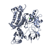
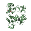
| 生物種 |  | ||||||
| 手法 |  X線回折 / シンクロトロン / 分子置換 / 解像度: 1.8 Å X線回折 / シンクロトロン / 分子置換 / 解像度: 1.8 Å | ||||||
 データ登録者 データ登録者 | Nagar, B. / Hantschel, O. / Young, M.A. / Scheffzek, K. / Veach, D. / Bornmann, W. / Clarkson, B. / Superti-Furga, G. / Kuriyan, J. | ||||||
 引用 引用 | ジャーナル: Cell(Cambridge,Mass.) / 年: 2003 タイトル: Structural basis for the autoinhibition of c-Abl tyrosine kinase 著者: Nagar, B. / Hantschel, O. / Young, M.A. / Scheffzek, K. / Veach, D. / Bornmann, W. / Clarkson, B. / Superti-Furga, G. / Kuriyan, J. #1: ジャーナル: Cell(Cambridge,Mass.) / 年: 2003タイトル: A myristoyl/phosphotyrosine switch regulates c-Abl 著者: Hantschel, O. / Nagar, B. / Guettler, S. / Kretzschmar, J. / Dorey, K. / Kuriyan, J. / Superti-Furga, G. | ||||||
| 履歴 |
| ||||||
| Remark 999 | SEQUENCE Numbering of the residues corresponds to the sequence database numbering of the isoform IV ...SEQUENCE Numbering of the residues corresponds to the sequence database numbering of the isoform IV of the protein. |
- 構造の表示
構造の表示
| 構造ビューア | 分子: MolmilJmol/JSmol |
|---|
- ダウンロードとリンク
-ダウンロード
| PDBx/mmCIF形式 | 1opk.cif.gz | 113.4 KB | 表示 | PDBx/mmCIF形式 |
|---|---|---|---|---|
| PDB形式 | pdb1opk.ent.gz | 84.3 KB | 表示 | PDB形式 |
| PDBx/mmJSON形式 | 1opk.json.gz | ツリー表示 | PDBx/mmJSON形式 | |
| その他 |  その他のダウンロード その他のダウンロード |
-検証レポート
| 文書・要旨 | 1opk_validation.pdf.gz | 729.5 KB | 表示 | wwPDB検証レポート |
|---|---|---|---|---|
| 文書・詳細版 | 1opk_full_validation.pdf.gz | 735.1 KB | 表示 | |
| XML形式データ | 1opk_validation.xml.gz | 21.3 KB | 表示 | |
| CIF形式データ | 1opk_validation.cif.gz | 31.3 KB | 表示 | |
| アーカイブディレクトリ | https://data.pdbj.org/pub/pdb/validation_reports/op/1opkftp://data.pdbj.org/pub/pdb/validation_reports/op/1opk | HTTPS FTP |
-関連構造データ


-リンク
 PDBj
PDBj


- 集合体
集合体
| 登録構造単位 | 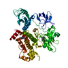
| ||||||||
|---|---|---|---|---|---|---|---|---|---|
| 1 |
| ||||||||
| 単位格子 |
|
-要素
| #1: タンパク質 | 分子量: 56189.164 Da / 分子数: 1 / 断片: SH3-SH2-kinase domain / 変異: D382N / 由来タイプ: 組換発現 / 由来: (組換発現) 発現宿主:   Spodoptera frugiperda (ツマジロクサヨトウ) Spodoptera frugiperda (ツマジロクサヨトウ)参照: UniProt: P00520, EC: 2.7.1.112 |
|---|---|
| #2: 化合物 | ChemComp-MYR /   分子量: 228.371 Da / 分子数: 1 / 由来タイプ: 合成 / 式: C14H28O2 分子量: 228.371 Da / 分子数: 1 / 由来タイプ: 合成 / 式: C14H28O2 |
| #3: 化合物 | ChemComp-P16 / 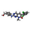  分子量: 427.283 Da / 分子数: 1 / 由来タイプ: 合成 / 式: C21H16Cl2N4O2 分子量: 427.283 Da / 分子数: 1 / 由来タイプ: 合成 / 式: C21H16Cl2N4O2 |
| #4: 化合物 | ChemComp-GOL /   分子量: 92.094 Da / 分子数: 1 / 由来タイプ: 合成 / 式: C3H8O3 分子量: 92.094 Da / 分子数: 1 / 由来タイプ: 合成 / 式: C3H8O3 |
| #5: 水 | ChemComp-HOH /  分子量: 18.015 Da / 分子数: 270 / 由来タイプ: 天然 / 式: H2O 分子量: 18.015 Da / 分子数: 270 / 由来タイプ: 天然 / 式: H2O |
-実験情報
-実験
| 実験 | 手法: X線回折 / 使用した結晶の数: 1 |
|---|
- 試料調製
試料調製
| 結晶 | マシュー密度: 2.49 Å3/Da / 溶媒含有率: 50.22 % | |||||||||||||||||||||||||||||||||||||||||||||||||
|---|---|---|---|---|---|---|---|---|---|---|---|---|---|---|---|---|---|---|---|---|---|---|---|---|---|---|---|---|---|---|---|---|---|---|---|---|---|---|---|---|---|---|---|---|---|---|---|---|---|---|
| 結晶化 | 温度: 293 K / 手法: 蒸気拡散法, ハンギングドロップ法 / pH: 7 詳細: 20% PEG 3350, 200 mM potassium nitrate, pH 7.0, VAPOR DIFFUSION, HANGING DROP, temperature 293K | |||||||||||||||||||||||||||||||||||||||||||||||||
| 結晶化 | *PLUS 温度: 20 ℃ / pH: 8 | |||||||||||||||||||||||||||||||||||||||||||||||||
| 溶液の組成 | *PLUS
|
-データ収集
| 回折 | 平均測定温度: 100 K |
|---|---|
| 放射光源 | 由来: シンクロトロン / サイト: ALS  / ビームライン: 8.2.2 / 波長: 1.0781 Å / ビームライン: 8.2.2 / 波長: 1.0781 Å |
| 検出器 | タイプ: ADSC QUANTUM 315 / 検出器: CCD / 日付: 2002年12月20日 / 詳細: mirrors |
| 放射 | モノクロメーター: Double crystal Si(111) / プロトコル: SINGLE WAVELENGTH / 単色(M)・ラウエ(L): M / 散乱光タイプ: x-ray |
| 放射波長 | 波長: 1.0781 Å / 相対比: 1 |
| 反射 | 解像度: 1.8→38 Å / Num. all: 50963 / Num. obs: 50963 / % possible obs: 99.9 % / Observed criterion σ(F): 0 / Observed criterion σ(I): -3 / 冗長度: 7.1 % / Biso Wilson estimate: 22 Å2 / Rsym value: 0.057 / Net I/σ(I): 29.6 |
| 反射 シェル | 解像度: 1.8→1.86 Å / 冗長度: 6.7 % / Mean I/σ(I) obs: 3.5 / Num. unique all: 5078 / Rsym value: 0.584 / % possible all: 100 |
| 反射 | *PLUS 最低解像度: 38 Å / Num. measured all: 560273 / Rmerge(I) obs: 0.057 |
| 反射 シェル | *PLUS 最高解像度: 1.8 Å / % possible obs: 100 % / Rmerge(I) obs: 0.584 |
- 解析
解析
| ソフトウェア |
| ||||||||||||||||||||||||||||||||||||
|---|---|---|---|---|---|---|---|---|---|---|---|---|---|---|---|---|---|---|---|---|---|---|---|---|---|---|---|---|---|---|---|---|---|---|---|---|---|
| 精密化 | 構造決定の手法: 分子置換 開始モデル: PDB ENTRIES 1M52, 2ABL 解像度: 1.8→37.59 Å / Rfactor Rfree error: 0.004 / Isotropic thermal model: RESTRAINED / 交差検証法: THROUGHOUT / σ(F): 0 / 立体化学のターゲット値: Engh & Huber
| ||||||||||||||||||||||||||||||||||||
| 溶媒の処理 | 溶媒モデル: FLAT MODEL / Bsol: 35.0282 Å2 / ksol: 0.366818 e/Å3 | ||||||||||||||||||||||||||||||||||||
| 原子変位パラメータ | Biso mean: 30.9 Å2
| ||||||||||||||||||||||||||||||||||||
| Refine analyze |
| ||||||||||||||||||||||||||||||||||||
| 精密化ステップ | サイクル: LAST / 解像度: 1.8→37.59 Å
| ||||||||||||||||||||||||||||||||||||
| 拘束条件 |
| ||||||||||||||||||||||||||||||||||||
| LS精密化 シェル | 解像度: 1.8→1.91 Å / Rfactor Rfree error: 0.013 / Total num. of bins used: 6
| ||||||||||||||||||||||||||||||||||||
| Xplor file |
| ||||||||||||||||||||||||||||||||||||
| 精密化 | *PLUS 最高解像度: 1.8 Å / 最低解像度: 38 Å | ||||||||||||||||||||||||||||||||||||
| 溶媒の処理 | *PLUS | ||||||||||||||||||||||||||||||||||||
| 原子変位パラメータ | *PLUS | ||||||||||||||||||||||||||||||||||||
| 拘束条件 | *PLUS
|
