 Movie
Movie Controller

[English] 日本語
 Yorodumi
- PDB-1nhz: Crystal Structure of the Antagonist Form of Glucocorticoid Receptor -
+ Open data

- Basic information
| Entry | Database: PDB / ID: 1nhz | ||||||
|---|---|---|---|---|---|---|---|
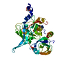
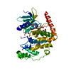
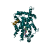
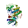
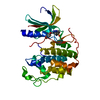
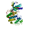
| Title | Crystal Structure of the Antagonist Form of Glucocorticoid Receptor | ||||||
 Components Components | GLUCOCORTICOID RECEPTOR | ||||||
 Keywords Keywords | Hormone receptor / PROTEIN-LIGAND COMPLEX / ANTI PARALLEL ALPHA HELIX SANDWICH | ||||||
| Function / homology |  Function and homology information Function and homology informationRegulation of NPAS4 gene transcription / regulation of glucocorticoid biosynthetic process / nuclear glucocorticoid receptor activity / steroid hormone binding / PTK6 Expression / glucocorticoid metabolic process / response to cortisol / neuroinflammatory response / mammary gland duct morphogenesis / microglia differentiation ...Regulation of NPAS4 gene transcription / regulation of glucocorticoid biosynthetic process / nuclear glucocorticoid receptor activity / steroid hormone binding / PTK6 Expression / glucocorticoid metabolic process / response to cortisol / neuroinflammatory response / mammary gland duct morphogenesis / microglia differentiation / astrocyte differentiation / maternal behavior / regulation of gluconeogenesis / adrenal gland development / cellular response to glucocorticoid stimulus / cellular response to steroid hormone stimulus / motor behavior / nuclear receptor-mediated steroid hormone signaling pathway / estrogen response element binding / cellular response to dexamethasone stimulus / FOXO-mediated transcription of oxidative stress, metabolic and neuronal genes / cellular response to transforming growth factor beta stimulus / core promoter sequence-specific DNA binding / steroid binding / HSP90 chaperone cycle for steroid hormone receptors (SHR) in the presence of ligand / TBP-class protein binding / RNA polymerase II transcription regulatory region sequence-specific DNA binding / SUMOylation of intracellular receptors / chromosome segregation / promoter-specific chromatin binding / synaptic transmission, glutamatergic / Hsp90 protein binding / Nuclear Receptor transcription pathway / response to wounding / positive regulation of miRNA transcription / DNA-binding transcription repressor activity, RNA polymerase II-specific / nuclear receptor activity / spindle / sequence-specific double-stranded DNA binding / Regulation of RUNX2 expression and activity / positive regulation of neuron apoptotic process / chromatin organization / DNA-binding transcription activator activity, RNA polymerase II-specific / gene expression / Potential therapeutics for SARS / DNA-binding transcription factor activity, RNA polymerase II-specific / nuclear speck / RNA polymerase II cis-regulatory region sequence-specific DNA binding / DNA-binding transcription factor activity / mitochondrial matrix / cell division / negative regulation of DNA-templated transcription / apoptotic process / regulation of transcription by RNA polymerase II / regulation of DNA-templated transcription / centrosome / synapse / protein kinase binding / chromatin / negative regulation of transcription by RNA polymerase II / signal transduction / positive regulation of transcription by RNA polymerase II / protein-containing complex / RNA binding / zinc ion binding / nucleoplasm / membrane / identical protein binding / nucleus / cytoplasm / cytosol Similarity search - Function | ||||||
| Biological species |  Homo sapiens (human) Homo sapiens (human) | ||||||
| Method |  X-RAY DIFFRACTION / SYNCHROTRON / MOLECULAR REPLACEMENT / Resolution: 2.3 Å X-RAY DIFFRACTION / SYNCHROTRON / MOLECULAR REPLACEMENT / Resolution: 2.3 Å | ||||||
 Authors Authors | Kauppi, B. / Jakob, C. / Farnegardh, M. / Yang, J. / Ahola, H. / Alarcon, M. / Calles, K. / Engstrom, O. / Harlan, J. / Muchmore, S. ...Kauppi, B. / Jakob, C. / Farnegardh, M. / Yang, J. / Ahola, H. / Alarcon, M. / Calles, K. / Engstrom, O. / Harlan, J. / Muchmore, S. / Ramqvist, A.-K. / Thorell, S. / Ohman, L. / Greer, J. / Gustafsson, J.-A. / Carlstedt-Duke, J. / Carlquist, M. | ||||||
 Citation Citation | Journal: J.Biol.Chem. / Year: 2003 Title: The Three-dimensional Structures of Antagonistic and Agonistic Forms of the Glucocorticoid Receptor Ligand-binding Domain: RU-486 INDUCES A TRANSCONFORMATION THAT LEADS TO ACTIVE ANTAGONISM. Authors: Kauppi, B. / Jakob, C. / Farnegardh, M. / Yang, J. / Ahola, H. / Alarcon, M. / Calles, K. / Engstrom, O. / Harlan, J. / Muchmore, S. / Ramqvist, A.-K. / Thorell, S. / Ohman, L. / Greer, J. / ...Authors: Kauppi, B. / Jakob, C. / Farnegardh, M. / Yang, J. / Ahola, H. / Alarcon, M. / Calles, K. / Engstrom, O. / Harlan, J. / Muchmore, S. / Ramqvist, A.-K. / Thorell, S. / Ohman, L. / Greer, J. / Gustafsson, J.-A. / Carlstedt-Duke, J. / Carlquist, M. | ||||||
| History |
|
- Structure visualization
| Structure viewer | Molecule: MolmilJmol/JSmol |
|---|
- Downloads & links
-Download
| PDBx/mmCIF format | 1nhz.cif.gz | 67.3 KB | Display | PDBx/mmCIF format |
|---|---|---|---|---|
| PDB format | pdb1nhz.ent.gz | 49 KB | Display | PDB format |
| PDBx/mmJSON format | 1nhz.json.gz | Tree view | PDBx/mmJSON format | |
| Others |  Other downloads Other downloads |
-Validation report
| Arichive directory | https://data.pdbj.org/pub/pdb/validation_reports/nh/1nhzftp://data.pdbj.org/pub/pdb/validation_reports/nh/1nhz | HTTPS FTP |
|---|
-Related structure data


-Links
 PDBj
PDBj


- Assembly
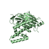
| Deposited unit | 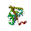
| ||||||||
|---|---|---|---|---|---|---|---|---|---|
| 1 |
| ||||||||
| 2 | 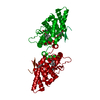
| ||||||||
| Unit cell |
| ||||||||
| Components on special symmetry positions |
|
-Components
| #1: Protein | Mass: 32155.072 Da / Num. of mol.: 1 Fragment: residue 500-777, hinge and steroid binding domains Mutation: N517D, F602S, C638D Source method: isolated from a genetically manipulated source Source: (gene. exp.) Homo sapiens (human) / Production host:   Spodoptera frugiperda (fall armyworm) / Strain (production host): Sf9 / References: UniProt: P04150 Spodoptera frugiperda (fall armyworm) / Strain (production host): Sf9 / References: UniProt: P04150 | ||
|---|---|---|---|
| #2: Chemical | ChemComp-486 /   Mass: 429.594 Da / Num. of mol.: 1 / Source method: obtained synthetically / Formula: C29H35NO2 Mass: 429.594 Da / Num. of mol.: 1 / Source method: obtained synthetically / Formula: C29H35NO2 | ||
| #3: Chemical |   Mass: 118.174 Da / Num. of mol.: 3 / Source method: obtained synthetically / Formula: C6H14O2 Mass: 118.174 Da / Num. of mol.: 3 / Source method: obtained synthetically / Formula: C6H14O2#4: Water | ChemComp-HOH / |  Mass: 18.015 Da / Num. of mol.: 128 / Source method: isolated from a natural source / Formula: H2O Mass: 18.015 Da / Num. of mol.: 128 / Source method: isolated from a natural source / Formula: H2O |
-Experimental details
-Experiment
| Experiment | Method: X-RAY DIFFRACTION / Number of used crystals: 1 |
|---|
- Sample preparation
| Crystal | Density Matthews: 2.7 Å3/Da / Density % sol: 54.04 % | |||||||||||||||||||||||||||||||||||||||||||||||||||||||||||||||
|---|---|---|---|---|---|---|---|---|---|---|---|---|---|---|---|---|---|---|---|---|---|---|---|---|---|---|---|---|---|---|---|---|---|---|---|---|---|---|---|---|---|---|---|---|---|---|---|---|---|---|---|---|---|---|---|---|---|---|---|---|---|---|---|---|
| Crystal grow | Temperature: 288 K / Method: vapor diffusion, hanging drop / pH: 8.2 Details: PEG 8000, 1, 6-hexanediol, NaSCN, Tris-HCl, pH 8.2, VAPOR DIFFUSION, HANGING DROP, temperature 288K | |||||||||||||||||||||||||||||||||||||||||||||||||||||||||||||||
| Crystal grow | *PLUS Temperature: 12 ℃ / pH: 8.5 / Method: vapor diffusion, hanging drop | |||||||||||||||||||||||||||||||||||||||||||||||||||||||||||||||
| Components of the solutions | *PLUS
|
-Data collection
| Diffraction | Mean temperature: 100 K |
|---|---|
| Diffraction source | Source: SYNCHROTRON / Site: ESRF  / Beamline: ID14-4 / Wavelength: 0.97 Å / Beamline: ID14-4 / Wavelength: 0.97 Å |
| Detector | Type: ADSC QUANTUM 4 / Detector: CCD / Date: Feb 26, 2002 |
| Radiation | Protocol: SINGLE WAVELENGTH / Monochromatic (M) / Laue (L): M / Scattering type: x-ray |
| Radiation wavelength | Wavelength: 0.97 Å / Relative weight: 1 |
| Reflection | Resolution: 2.3→39.26 Å / Num. all: 14833 / Num. obs: 14815 / % possible obs: 99.1 % / Observed criterion σ(F): 0 / Observed criterion σ(I): 0 / Redundancy: 3.5 % / Biso Wilson estimate: 35 Å2 / Rmerge(I) obs: 0.045 / Rsym value: 0.035 / Net I/σ(I): 8.8 |
| Reflection shell | Resolution: 2.3→2.42 Å / Redundancy: 3.7 % / Rmerge(I) obs: 0.141 / Mean I/σ(I) obs: 6.1 / Num. unique all: 2064 / Rsym value: 0.121 / % possible all: 99.1 |
| Reflection | *PLUS Highest resolution: 2.3 Å / Lowest resolution: 40 Å / % possible obs: 98.1 % / Rmerge(I) obs: 0.106 |
| Reflection shell | *PLUS % possible obs: 98 % / Rmerge(I) obs: 0.348 / Mean I/σ(I) obs: 2.1 |
- Processing
| Software |
| ||||||||||||||||||||||||||||||||||||||||||||||||||||||||||||||||||||||||||||||||||||||||||||||||||||||||||||||||||||||||||||||||||
|---|---|---|---|---|---|---|---|---|---|---|---|---|---|---|---|---|---|---|---|---|---|---|---|---|---|---|---|---|---|---|---|---|---|---|---|---|---|---|---|---|---|---|---|---|---|---|---|---|---|---|---|---|---|---|---|---|---|---|---|---|---|---|---|---|---|---|---|---|---|---|---|---|---|---|---|---|---|---|---|---|---|---|---|---|---|---|---|---|---|---|---|---|---|---|---|---|---|---|---|---|---|---|---|---|---|---|---|---|---|---|---|---|---|---|---|---|---|---|---|---|---|---|---|---|---|---|---|---|---|---|---|
| Refinement | Method to determine structure: MOLECULAR REPLACEMENT / Resolution: 2.3→62.02 Å / Cor.coef. Fo:Fc: 0.927 / Cor.coef. Fo:Fc free: 0.881 / SU B: 6.11 / SU ML: 0.156 / Cross valid method: THROUGHOUT / σ(F): 0 / ESU R: 0.299 / ESU R Free: 0.253 / Stereochemistry target values: MAXIMUM LIKELIHOOD / Details: HYDROGENS HAVE BEEN ADDED IN THE RIDING POSITIONS
| ||||||||||||||||||||||||||||||||||||||||||||||||||||||||||||||||||||||||||||||||||||||||||||||||||||||||||||||||||||||||||||||||||
| Solvent computation | Ion probe radii: 0.8 Å / Shrinkage radii: 0.8 Å / VDW probe radii: 1.4 Å / Solvent model: BABINET MODEL WITH MASK | ||||||||||||||||||||||||||||||||||||||||||||||||||||||||||||||||||||||||||||||||||||||||||||||||||||||||||||||||||||||||||||||||||
| Displacement parameters | Biso mean: 16.433 Å2
| ||||||||||||||||||||||||||||||||||||||||||||||||||||||||||||||||||||||||||||||||||||||||||||||||||||||||||||||||||||||||||||||||||
| Refinement step | Cycle: LAST / Resolution: 2.3→62.02 Å
| ||||||||||||||||||||||||||||||||||||||||||||||||||||||||||||||||||||||||||||||||||||||||||||||||||||||||||||||||||||||||||||||||||
| Refine LS restraints |
| ||||||||||||||||||||||||||||||||||||||||||||||||||||||||||||||||||||||||||||||||||||||||||||||||||||||||||||||||||||||||||||||||||
| LS refinement shell | Resolution: 2.3→2.36 Å / Total num. of bins used: 20 /
| ||||||||||||||||||||||||||||||||||||||||||||||||||||||||||||||||||||||||||||||||||||||||||||||||||||||||||||||||||||||||||||||||||
| Refinement | *PLUS Highest resolution: 2.3 Å / Lowest resolution: 40 Å / Rfactor Rfree: 0.263 / Rfactor Rwork: 0.228 | ||||||||||||||||||||||||||||||||||||||||||||||||||||||||||||||||||||||||||||||||||||||||||||||||||||||||||||||||||||||||||||||||||
| Solvent computation | *PLUS | ||||||||||||||||||||||||||||||||||||||||||||||||||||||||||||||||||||||||||||||||||||||||||||||||||||||||||||||||||||||||||||||||||
| Displacement parameters | *PLUS | ||||||||||||||||||||||||||||||||||||||||||||||||||||||||||||||||||||||||||||||||||||||||||||||||||||||||||||||||||||||||||||||||||
| LS refinement shell | *PLUS Highest resolution: 2.3 Å / Lowest resolution: 2.36 Å |
